Reticulospinal tract
| Reticulospinal tract | |
|---|---|
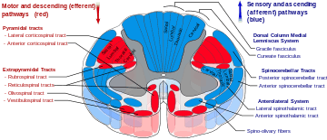 Reticulospinal tract is labeled in red, near-center in figure (text tag at left). | |
| Identifiers | |
| NeuroNames | hier-802 |
| NeuroLex ID | Reticulospinal tract |
| TA | A14.1.04.124 |
| FMA | 72639 |
The reticulospinal tract (or anterior reticulospinal tract) is an extrapyramidal motor tract that descends from the reticular formation in two tracts to act on the motor neurons supplying the trunk and proximal limb muscles. It is involved mainly in locomotion and postural control, although it does have other effects as well.[1]
Functions
1. Integrates information from the motor systems to coordinate automatic movements of locomotion and posture
2. Facilitates and inhibits voluntary movement; influences muscle tone
3. Mediates autonomic functions
4. Modulates pain impulses
5. Influences blood flow to lateral geniculate nucleus of the thalamus.
Components
The tract is divided into two parts, the medial (or pontine) and lateral (or medullary) reticulospinal tracts (MRST and LRST).
- The MRST is responsible for exciting anti-gravity, extensor muscles. The fibers of this tract arise from the caudal pontine reticular nucleus and the oral pontine reticular nucleus and project to the lamina VII and lamina VIII of the spinal cord (BrainInfo)
- The LRST is responsible for inhibiting excitatory axial extensor muscles of movement. The fibers of this tract arise from the medullary reticular formation, mostly from the gigantocellular nucleus, and descend the length of the spinal cord in the anterior part of the lateral column. The tract terminates in lamina VII mostly with some fibers terminating in lamina IX of the spinal cord.
The sensory tract conveying information in the opposite direction is known as the "spinoreticular tract".
Clinical significance
Mostly inhibited by the corticospinal tract, if damage occurs at the level of or below the red nucleus (e.g. to the superior colliculus) it is called decerebration and causes decerebrate rigidity, an unopposed extension of the head and limbs.
The reticulospinal tracts also provide a pathway by which the hypothalamus can control sympathetic thoracolumbar outflow and parasympathetic sacral outflow.
See also
References
- ↑ FITGERALD, M J Turlough (2012). Clinical Neuroanatomy and Neuroscience. Philadelphia: Saunders Elsevier. p. 192. ISBN 978-0-7020-3738-2.
External links
- BrainInfo: reticulospinal tract, pontine reticulospinal tract, and medullary reticulospinal tract.
- http://www.lib.mcg.edu/edu/eshuphysio/program/section8/8ch6/s8ch6_29.htm
- http://www.mona.uwi.edu/fpas/courses/physiology/neurophysiology/The%20Reticulospinal%20Pathway.htm