Reticular formation
| Reticular formation | |
|---|---|
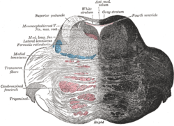 Axial section of the pons, at its upper part. (Formatio reticularis labeled at left.) | |
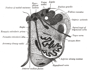 Section of the medulla oblongata at about the middle of the olive. (Formatio reticularis grisea and formatio reticularis alba labeled at left.) | |
| Details | |
| Identifiers | |
| Latin | formatio reticularis |
| MeSH | A08.186.211.132.772 |
| NeuroNames | ancil-225 |
| NeuroLex ID | Reticular formation |
| TA |
A14.1.00.021 A14.1.05.403 A14.1.06.327 |
| FMA | 77719 |
The reticular formation is a set of interconnected nuclei that are located throughout the brainstem. The reticular formation is not anatomically well defined because it includes neurons located in diverse parts of the brain. The neurons of the reticular formation all play a crucial role in maintaining behavioral arousal and consciousness. The functions of the reticular formation are modulatory and premotor. The modulatory functions are primarily found in the rostral sector of the reticular formation and the premotor functions are localized in the neurons in more caudal regions.
The reticular formation is divided into three columns: raphe nuclei (median), gigantocellular reticular nuclei (medial zone), and parvocellular reticular nuclei (lateral zone). The raphe nuclei are the place of synthesis of the neurotransmitter serotonin, which plays an important role in mood regulation. The gigantocellular nuclei are involved in motor coordination. The parvocellular nuclei regulate exhalation.[1]
It is essential for governing some of the basic functions of higher organisms and is one of the phylogenetically oldest portions of the brain.
Structure

The reticular formation has been functionally cleaved both sagittally and coronally.
Traditionally the nuclei are divided into three columns
- In the median column – the raphe nuclei
- In the medial column – gigantocellular nuclei (because of larger size of the cells)
- In the lateral column – parvocellular nuclei (because of smaller size of the cells)
The original functional differentiation was a division of caudal and rostral, this was based upon the observation that the lesioning of the rostral reticular formation induces a hypersomnia in the cat brain. In contrast, lesioning of the more caudal portion of the reticular formation produces insomnia in cats. This study has led to the idea that the caudal portion inhibits the rostral portion of the reticular formation.
Sagittal division reveals more morphological distinctions. The raphe nuclei form a ridge in the middle of the reticular formation, and, directly to its periphery, there is a division called the medial reticular formation. The medial RF is large and has long ascending and descending fibers, and is surrounded by the lateral reticular formation. The lateral RF is close to the motor nuclei of the cranial nerves, and mostly mediates their function.
Medial and lateral reticular formation
The medial reticular formation and lateral reticular formation are two columns of neuronal nuclei with ill-defined boundaries that send projections through the medulla and into the mesencephalon (midbrain). The nuclei can be differentiated by function, cell type, and projections of efferent or afferent nerves.
Function
The reticular formation consists of more than 100 small neural networks, with varied functions including the following:
- Somatic motor control – Some motor neurons send their axons to the reticular formation nuclei, giving rise to the reticulospinal tracts of the spinal cord. These tracts function in maintaining tone, balance, and posture—especially during body movements. The reticular formation also relays eye and ear signals to the cerebellum so that the cerebellum can integrate visual, auditory, and vestibular stimuli in motor coordination. Other motor nuclei include gaze centers, which enable the eyes to track and fixate objects, and central pattern generators, which produce rhythmic signals to the muscles of breathing and swallowing.
- Cardiovascular control – The reticular formation includes the cardiac and vasomotor centers of the medulla oblongata.
- Pain modulation – The reticular formation is one means by which pain signals from the lower body reach the cerebral cortex. It is also the origin of the descending analgesic pathways. The nerve fibers in these pathways act in the spinal cord to block the transmission of some pain signals to the brain.
- Sleep and consciousness – The reticular formation has projections to the thalamus and cerebral cortex that allow it to exert some control over which sensory signals reach the cerebrum and come to our conscious attention. It plays a central role in states of consciousness like alertness and sleep. Injury to the reticular formation can result in irreversible coma.
- Habituation – This is a process in which the brain learns to ignore repetitive, meaningless stimuli while remaining sensitive to others. A good example of this is a person who can sleep through loud traffic in a large city, but is awakened promptly due to the sound of an alarm or crying baby. Reticular formation nuclei that modulate activity of the cerebral cortex are part of the reticular activating system.[2][3]
Clinical significance
Mass lesions in the brainstem cause severe alterations in level of consciousness (such as coma) because of their effects on the reticular formation.[4] Bilateral damage to the reticular formation of the midbrain may lead to coma or death.[5]
Lesions in the reticular formation have been found in the brains of people who have post-polio syndrome, and some imaging studies have shown abnormal activity in this area in people with chronic fatigue syndrome, indicating a high likelihood that damage to the reticular formation is responsible for the fatigue associated with these syndromes.
History
The term "reticular formation" was coined in the late 19th century, coinciding with Ramon y Cajal’s neuron doctrine. Allan Hobson states in his book The Reticular Formation Revisited that the name is an etymological vestige from the fallen era of the aggregate field theory in the neural sciences. The term "reticulum" means "netlike structure," which is what the reticular formation resembles at first glance. It has been described as being either too complex to study or an undifferentiated part of the brain with no organization at all. Eric Kandel describes the reticular formation as being organized in a similar manner to the intermediate gray matter of the spinal cord. This chaotic, loose, and intricate form of organization is what has turned off many researchers from looking farther into this particular area of the brain. The cells lack clear ganglionic boundaries, but do have clear functional organizations and distinct cell types.
The term "reticular formation" is seldom used anymore except to speak in generalities. Modern scientists usually refer to the individual nuclei that comprise the reticular formation.
See also
- Locus coeruleus
- Pedunculopontine nucleus
- Medial pontine reticular formation
- Reticular activating system
- Midbrain reticular formation
References
- ↑ "The Brain From Top To Bottom". Thebrain.mcgill.ca. Retrieved 2016-04-28.
- ↑ "Anatomy of the Brain - Reticular Formation". Biology.about.com. 2015-07-07. Retrieved 2016-04-28.
- ↑ Saladin, Kenneth S. Anatomy & Physiology the Unity of Form and Function. Dubuque: McGraw-Hill, 2009. Print.
- ↑ Tindall SC (1990). "Level of consciousness". In Walker HK, Hall WD, Hurst JW. Clinical Methods: The History, Physical, and Laboratory Examinations. Butterworth Publishers. Retrieved 2008-07-04.
- ↑ The Human Brain: An Introduction to its Functional Anatomy 5th ed by J Nolte chpt 11 pp. 262–290
Additional images
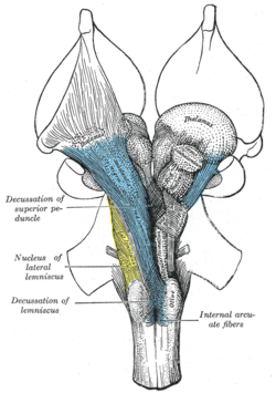 Deep dissection of brain-stem. Ventral view.
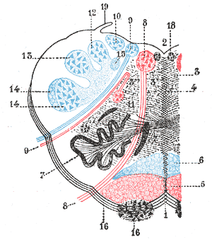 The formatio reticularis of the medulla oblongata, shown by a transverse section passing through the middle of the olive.
External links
| Look up reticular formation in Wiktionary, the free dictionary. |