Corticopontine fibers
| Corticopontine fibers | |
|---|---|
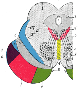 Coronal section through mid-brain. 1. Corpora quadrigemina. 2. Cerebral aqueduct. 3. Central gray stratum. 4. Interpeduncular space. 5. Sulcus lateralis. 6. Substantia nigra. 7. Red nucleus of tegmentum. 8. Oculomotor nerve, with 8’, its nucleus of origin. a. Lemniscus (in blue) with a’ the medial lemniscus and a" the lateral lemniscus. b. Medial longitudinal fasciculus. c. Raphé. d. Temporopontine fibers. e. Portion of medial lemniscus, which runs to the lentiform nucleus and insula. f. Cerebrospinal fibers. g. Frontopontine fibers. | |
| Details | |
| Identifiers | |
| Latin | fibrae corticopontinae, tractus corticopontinus |
| NeuroNames | ancil-375 |
| TA | A14.1.05.107 |
| FMA | 75190 |
Corticopontine fibers [1] are projections from the cerebral cortex to the pontine nuclei.[2]
Depending upon the lobe of origin, they can be classified as frontopontine fibers, parietopontine fibers, temporopontine fibers or occipitopontine fibers.[3]
References
- ↑ Kamali A, Kramer LA, Frye RE, Butler IJ, Hasan KM. Diffusion tensor tractography of the human brain cortico-ponto-cerebellar pathways: a quantitative preliminary study. J Magn Reson Imaging. 2010 Oct;32(4):809-17. doi: 10.1002/jmri.22330. PMID 20882611
- ↑ Leergaard TB, Bjaalie JG (November 2007). "Topography of the complete corticopontine projection: From experiments to principal Maps". Front Neurosci. 1 (1): 211–23. doi:10.3389/neuro.01.1.1.016.2007. PMC 2518056
 . PMID 18982130.
 - ↑ http://braininfo.rprc.washington.edu/AncilDefinition.aspx?ID=1322&questID=1322
External links
This article is issued from Wikipedia - version of the 3/23/2015. The text is available under the Creative Commons Attribution/Share Alike but additional terms may apply for the media files.