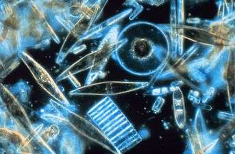
Picoplankton
Picoplankton is the fraction of plankton composed by cells between 0.2 and 2 μm that can be either prokaryotic and eukaryotic phototrophs and heterotrophs:
- photosynthetic Main article: Photosynthetic picoplankton
- heterotrophic Main article: Heterotrophic picoplankton
They are prevalent amongst microbial plankton communities of both freshwater and marine ecosystems. They have an important role in making up a significant portion of the total biomass of phytoplankton communities
Classification
In general, plankton can be categorized on the basis of physiological, taxonomic, or dimensional characteristics. Subsequently, a generic classification of a plankton includes:
However, there is a simpler scheme that categorizes plankton based on a logarithmic size scale:
- Macroplankton (200-2000 μm)
- Micro-plankton (20-200 μm)
- Nanoplankton (2-20 μm)
This was even further expanded to include picoplankton (0.2-2 μm) and fem-toplankton (0.02-0.2 μm), as well as net plankton, ultraplankton. Now that picoplankton have been characterized, they have their own further subdivisions such as prokaryotic and eukaryotic phototrophs and heterotrophs that are spread throughout the world in various types of lakes and tropic states. In order to differentiate between autotrophic picoplankton and heterotrophic picoplankton, the autotrophs could have photosynthetic pigments and the ability to show autofluorescence, which would allow for their enumeration under epifluorescence microscopy. This is how minute eukaryotes first became known.[1] Overall, picoplankton play an essential role in oligotrophic dimicitc lakes because they are able to produce and then accordingly recycle dissolved organic matter (DOM) in a very efficient manner under circumstance when competition of other phytoplankters is disturbed by factors such as limiting nutrients and predators. Picoplankton are responsible for the most primary productivity in oligotrophic gyres, and are distinguished from nanoplankton and microplankton.[2] Because they are small, they have a greater surface to volume ratio, enabling them to obtain the scarce nutrients in these ecosystems. Furthermore, some species can also be mixotrophic. The smallest of cells (200 nm) are on the order of nanometers, not picometers. The SI prefix pico- is used quite loosely here, as nanoplankton and microplankton are only 10 and 100 times larger, respectively, although it is somewhat more accurate when considering the volume rather than the length.
Measurement
Marine scientists have slowly begun to understand in the last 10 or 15 years the importance of even the smallest subdivisions of plankton and their role in aquatic food webs and in organic and inorganic nutrient recycling. Therefore, being able to accurately measure the biomass and size distribution of picoplankton communities has now become rather essential. Two of the prevalent methods used to identify and enumerate picoplankton are fluorescence microscopy and visual counting. However, both methods are outdated because of their time-consuming and inaccurate nature. As a result, newer, faster, and more accurate methods have emerged lately, including flow cytometry and image-analyzed fluorescence microscopy. Both techniques are efficient in measuring nano plankton and auto-fluorescing phototrophic picoplankton. However, measuring very minute size ranges of picoplankton has often proven to be difficult to measure, which is why Charge-coupled devices (CCD) and video cameras are now being used to measure small picoplankton, although a slow-scan CCD-based camera is more effective at detecting and sizing tiny particles such as bacteria that is fluorochrome-stained.
See also
References
- ↑ C. Callieri and J. G. Stockner, Freshwater autotrophic picoplankton: a review, J. Limnol., 2002, 61, 1–14
- ↑ Vershinin, Alexander. "Phytoplankton in the Black Sea". Russian Federal Children Center Orlyonok.
- ↑ Viles, C L, and M E Sieracki. “Measurement of Marine Picoplankton Cell Size by Using a Cooled, Charge-Coupled Device Camera with Image-Analyzed Fluorescence Microscopy.” Applied and Environmental Microbiology 58.2 (1992): 584–592. Print.