Occipital bone
| Occipital bone | |
|---|---|
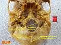
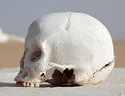 Human skull (Occipital bone is at bottom right). | |
 Position of occipital bone (shown in green) | |
| Details | |
| Articulations | the two parietals, the two temporals, the sphenoid, and the atlas |
| Identifiers | |
| Latin | os occipitale |
| TA | A02.1.04.001 |
| FMA | 52735 |
The occipital bone (/ˌɒkˈsɪpᵻtəl/), is a cranial dermal bone, and is the main bone of the occiput (back and lower part of the skull). It is trapezoidal in shape and curved on itself like a shallow dish. The occipital bone overlies the occipital lobes of the cerebrum. At the base of the skull in the occipital bone there is a large oval opening called the foramen magnum, which allows the passage of the spinal cord.
Like the other cranial bones it is classed as a flat bone. Due to its many attachments and features the occipital bone is described in terms of separate parts. From its front to the back is the basilar part, at the sides of the foramen magnum are the lateral parts, and the back is named as the squamous part.
The basilar part is a thick, somewhat quadrilateral piece in front of the foramen magnum and directed towards the pharynx. This is also called the basioccipital.
The lateral parts of the occipital bone are on either side of the foramen magnum and these are also called the exoccipitals.
The squamous part is the curved, expanded plate behind the foramen magnum and is the largest part of the occipital bone.
Structure
The occipital bone, like the other cranial bones has outer and inner layers (also called plates or tables) of cortical bone tissue between which is the cancellous bone tissue or diploë; the bone is especially thick at the ridges, protuberances, condyles, and anterior part of the basilar part; in the inferior cerebellar fossae it is thin, semitransparent, and without diploë.
Outer surface

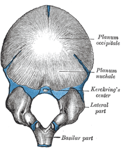
Near the middle of the outer surface of the squamous part of the occipital (the largest part) there is a prominence – the external occipital protuberance. The highest point of this is called the inion.
From the inion, along the midline of the squamous part until the foramen magnum, runs a ridge – the external occipital crest (also called the medial nuchal line) and this gives attachment to the nuchal ligament.
Running across the outside of the occipital bone are three curved lines and one line (the medial line) that runs down to the foramen magnum. These are known as the nuchal lines which give attachment to various ligaments and muscles. They are named as the highest, superior and inferior nuchal lines. The inferior nuchal line runs across the mid-point of the medial nuchal line. The area above the highest nuchal line is termed the occipital plane and the area below this line is termed the nuchal plane.
Inner surface

The inner surface of the occipital bone forms the base of the posterior cranial fossa. The foramen magnum is a large hole situated in the middle, with the clivus, a smooth part of the occipital bone travelling upwards in front of it. The median internal occipital crest travels behind it to the internal occipital protuberance, and serves as a point of attachment to the falx cerebri. To the sides of the foramen sitting at the junction between the lateral and base of the occipital bone are the hypoglossal canals. Further out, at each junction between the occipital and petrous part of the temporal bone lies a jugular foramen.[1]
The inner surface of the occipital bone is marked by dividing lines as shallow ridges, that form four fossae or depressions. The lines are called the cruciform (cross-shaped) eminence. At the mid-point where the lines intersect a raised part is formed called the internal occipital protuberance. From each side of this eminence runs a groove for the transverse sinuses.
There are two midline skull landmarks at the foramen magnum. The basion is the most anterior point of the opening and the opisthion is the point on the opposite posterior part. The basion lines up with the dens.
Foramen magnum
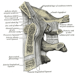
The foramen magnum (Latin: large hole) is a large oval foramen longest front to back; it is wider behind than in front where it is encroached upon by the occipital condyles. The clivus, a smooth bony section, travels upwards on the front surface of the foramen, and the median internal occipital crest travels behind it.[2]
Through the foramen passes the medulla oblongata and its membranes, the accessory nerves, the vertebral arteries, the anterior and posterior spinal arteries, and the tectorial membrane and alar ligaments.
Angles
The superior angle of the occipital bone articulates with the occipital angles of the parietal bones and, in the fetal skull, corresponds in position with the posterior fontanelle.
The inferior angle is fused with the body of the sphenoid. The lateral angles are situated at the extremities of the groove for the transverse sinuses: each is received into the interval between the mastoid angle of the parietal and the mastoid part of the temporal.
Borders
The superior borders extend from the superior to the lateral angles: they are deeply serrated for articulation with the occipital borders of the parietals, and form by this union the lambdoidal suture.
The inferior borders extend from the lateral angles to the inferior angle; the upper half of each articulates with the mastoid portion of the corresponding temporal, the lower half with the petrous part of the same bone.
These two portions of the inferior border are separated from one another by the jugular process, the notch on the anterior surface of which forms the posterior part of the jugular foramen.
-

Lambdoidal suture. It separates occipital bone from parietal bones.
-

Occipitomastoid suture. It separates occipital bone and mastoid portion of the temporal bone.
Development
The occipital plane [Fig. 3] of the squamous part of the occipital bone is developed in membrane, and may remain separate throughout life when it constitutes the interparietal bone; the rest of the bone is developed in cartilage.
The number of nuclei for the occipital plane is usually given as four, two appearing near the middle line about the second month, and two some little distance from the middle line about the third month of fetal life.
The nuchal plane of the squamous part is ossified from two centers, which appear about the seventh week of fetal life and soon unite to form a single piece.
Union of the upper and lower portions of the squamous part takes place in the third month of fetal life.
An occasional centre (Kerckring) appears in the posterior margin of the foramen magnum during the fifth month; this forms a separate ossicle (sometimes double) which unites with the rest of the squamous part before birth.
Each of the lateral parts begins to ossify from a single center during the eighth week of fetal life. The basilar portion is ossified from two centers, one in front of the other; these appear about the sixth week of fetal life and rapidly coalesce.
The occipital plane is said to be ossified from two centers and the basilar portion from one.
About the fourth year the squamous part and the two lateral parts unite, and by about the sixth year the bone consists of a single piece. Between the 18th and 25th years the occipital and sphenoid bone become united, forming a single bone.
Clinical significance
Trauma to the occiput can cause a fracture of the base of the skull, called a basilar skull fracture.
Genetic disorders can cause a prominent occiput as found in Edwards syndrome, and Beckwith–Wiedemann syndrome.
The identification of the location of the fetal occiput is important in delivery.
Etymology
Occipital : from Latin occiput "back of the skull," from ob "against, behind" + caput "head" . distinguished from sinciput (anterior part of the skull).[3]
Other animals
In many animals these parts stay separate throughout life; for example, in the dog as four parts: squamous part (supraoccipita); lateral parts–left and right parts (exoccipital); basilar part (basioccipital).
The occipital bone is part of the endocranium, the most basal portion of the skull. In Chondrichthyes and Agnathans, the occipital does not form as a separate element, but remains part of the chondrocranium throughout life. In most higher vertebrates, the foramen magnum is surrounded by a ring of four bones. The basioccipital lies in front of the opening, the two exoccipital condyles lie to either side, and the larger supraoccipital lies to the posterior, and forms at least part of the rear of the cranium. In many bony fish and amphibians, the supraoccipital is never ossified, and remains as cartilage throughout life. In primitive forms the basioccipital and exoccipitals somewhat resemble the centrum and neural arches of a vertebra, and form in a similar manner in the embryo. Together, these latter bones usually form a single concave circular condyle for the articulation of the first vertebra.[4]
In mammals, however, the condyle has divided in two, a pattern otherwise seen only in a few amphibians. Most mammals also have a single fused occipital bone, formed from the four separate elements around the foramen magnum, along with the paired postparietal bones that form the rear of the cranial roof in other vertebrates.[4]
Additional images
-

Position of occipital bone (shown in green). Animation.
-

Outer surface.
-

Inner surface. Frontal bone and parietal bones are removed.
-

Occipital bone
-
Occipital bone
-
Median sagittal section through the occipital bone and first three cervical vertebræ.
-
Basilar part
-

Occipital bone
See also
| Wikimedia Commons has media related to Occipital bones. |
References
This article incorporates text in the public domain from the 20th edition of Gray's Anatomy (1918)
- ↑ Gray's Anatomy 2008, p. 424-425.
- ↑ Gray's Anatomy 2008, p. 425.
- ↑ "occipital" A Dictionary of Zoology. Ed. Michael Allaby. Oxford University Press 2009
- 1 2 Romer, Alfred Sherwood; Parsons, Thomas S. (1977). The Vertebrate Body. Philadelphia, PA: Holt-Saunders International. pp. 221–244. ISBN 0-03-910284-X.
- Books
- editor-in-chief, Susan Standring ; section editors, Neil R. Borley; et al. (2008). Gray's anatomy : the anatomical basis of clinical practice (40th ed.). London: Churchill Livingstone. ISBN 978-0-8089-2371-8.