Ethmoid sinus
| Ethmoidal sinus | |
|---|---|
 | |
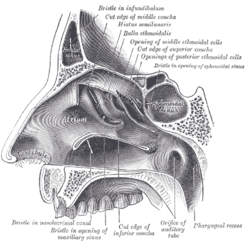
 Coronal section of nasal cavities. | |
| Details | |
| Nerve | posterior ethmoidal nerve |
| Identifiers | |
| Latin |
Cellulae ethmoidales, labyrinthi ethmoidales |
| MeSH | A04.531.621.267 |
| TA | A06.1.03.005 |
| FMA | 71958 |
The ethmoidal sinuses or ethmoidal air cells of the ethmoid bone are one of the four paired paranasal sinuses. They are a variable in both size and number of small cavities in the lateral mass of each of the ethmoid bones and cannot be palpated during an extraoral examination.[1] They are divided into the anterior, middle and posterior groups (see below). The ethmoidal air cells consist of numerous thin-walled cavities situated in the ethmoidal labyrinth and completed by the frontal, maxilla, lacrimal, sphenoidal, and palatine bones. They lie between the upper parts of the nasal cavities and the orbits, and are separated from these cavities by thin bony laminae.[2]
Groups of sinuses
The groups of the ethmoidal cells are air cells:[2]
- The posterior group (sometimes the posterior ethmoidal sinus) drains into the superior meatus above the middle nasal concha; sometimes one or more opens into the sphenoidal sinus.
- The middle group (sometimes the middle ethmoidal sinus) drains into the middle meatus of the nose on or above the bulla ethmoidalis.
- The anterior group (sometimes the anterior ethmoidal sinus) drains into the middle meatus of the nose by way of the infundibulum.
Development
The ethmoidal cells (sinuses) are present at birth, however by 2 years of age they are recognisable through the use of Computerised Tomography (CT) scanning.[3]
Innervation
The ethmoidal air cells receive sensory fibers from the anterior and posterior ethmoidal nerves, and the orbital branches of the pterygopalatine ganglion, which carry the postganglionic parasympathetic nerve fibers for mucous secretion from the facial nerve.
Haller cell
Haller cells are infraorbital ethmoidal air cells lateral to the lamina papyracea. These may arise from the anterior or posterior ethmoidal sinuses.
Pathology
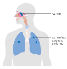

Acute ethmoiditis in childhood and ethmoidal carcinoma may spread superiorly causing meningitis and cerebrospinal fluid leakage or it may spread laterally into the orbit causing proptosis and diplopia.[4]
Additional images
-

Ethmoid bone from the right side.
-

Left orbicularis oculi, seen from behind.
-
Lateral wall of nasal cavity; the three nasal conchæ have been removed.
-

Paranasal sinuses
-

Ethmoid cells
-

Ethmoid sinus. Ethmoidal air cells.Deep dissection. Superior view.
References
This article incorporates text in the public domain from the 20th edition of Gray's Anatomy (1918)
External links
- Anatomy figure: 33:04-07 at Human Anatomy Online, SUNY Downstate Medical Center
- Anatomy photo:33:st-0711 at the SUNY Downstate Medical Center