Malum perforans
| Malum perforans | |
|---|---|
 | |
| Diabetic foot ulcer | |
| Classification and external resources | |
| Specialty | dermatology |
| ICD-10 | L97 (ILDS L97) |
Malum perforans (also known as neurotrophic ulcer and trophic ulcer) is a long-lasting, usually painless ulcer that penetrates deep into or through the skin, usually on the sole of the foot (in which case it may be called malum perforans pedis). It is often a complication in diabetes mellitus and other conditions affecting the nerves.[1]
Cause
This condition results from denervation of areas exposed to day-to-day friction of bony prominences. The denervation may be result of any of the following diseases:
- Spinal injuries
- Leprosy
- Peripheral nerve injury
- Diabetic neuropathy
- Tabes dorsalis
- Transverse myelitis
- Meningomyelocele
Pathophysiology
Normal pressure and pain sensations are essential for protecting the foot from excessive and prolonged pressures over bony prominences. In insensitive foot, such as in diabetic neuropathy, soft tissues are exposed to excessive pressures without knowledge of the individual. In other words, by nerve damage in the feet, the patients get no feedback on the impact of the feet when walking. These ulcers start with callosity under which suppuration takes place. The pus comes out and a hole forms under which the lesion grows deeper. This leads to punched-out, painless ulcers usually under metatarsal heads, tip of toe, proximal interphalageal joint of a hammertoe or on the heel.[2] In non-ambulatory patients, these ulcers are found on buttocks and back of heel.
Characteristics
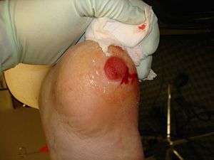
These ulcers have punched-out edge and slough in floor, resembling gummatous ulcer. Surrounding area might have loss of sensation.
Diagnosis
Diagnosis is clinical. Sensation is tested using graded monofilament.[2]
Treatment
Underlying cause of neuropathy is first treated. Necrotic portions of the wound are removed and wound is kept moist at associations. Infected ulcers are administered antibiotics. Skin grafting is one of the options. It has been shown that ultrasound may increase the acceptance of graft at trophic ulcer sites.[3]
See also
References
- ↑ Siebel, R; Baumgartner, R; Greitemann, B; Junker, Th. (6 December 2012). "The Treatment of Malum Perforans Pedis". In Altmeyer, Peter; Hoffmann, Klaus; el Gammal, Stephan; et al. Wound Healing and Skin Physiology. Springer Science & Business Media. p. 423. ISBN 978-3-642-77882-7.
- 1 2 DeGowin's Diagnostic Examination 8th ed. 2004. p. 157. ISBN 9780071409230.
- ↑ Physical principles of medical ultrasonics. 2004. p. 421. ISBN 9780471970026.