Brown tumor
| Brown tumor | |
|---|---|
|
Brown tumours of the hands in a patient with hyperparathyroidism. | |
| Classification and external resources | |
| ICD-9-CM | 252.01 |
| DiseasesDB | 30721 |
| MeSH | D010002 |
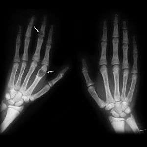
The brown tumor is a bone lesion that arises in settings of excess osteoclast activity, such as hyperparathyroidism. It is not a true neoplasm, as the term "tumor" suggests; however, it may mimic a true neoplasm.[1] Brown tumours are radiolucent on x-ray.
Pathology

Brown tumours consist of fibrous tissue, woven bone and supporting vasculature, but no matrix. The osteoclasts consume the trabecular bone that osteoblasts lay down and this front of reparative bone deposition followed by additional resorption can expand beyond the usual shape of the bone, involving the periosteum thus causing bone pain. The characteristic brown coloration results from hemosiderin deposition into the osteolytic cysts. Hemosiderin deposition is not a distinctive feature of brown tumors; it may also be seen in giant cell tumors of the bone.[2][3]
Brown tumors may be rarely associated with ectopic parathyroid adenomas.[4]
Epidemiology
Age and gender have an effect on the incidence of these lesions; they are more prevalent in women than men (though still common in both genders), and they appear more frequently with age. Due to the standard of medical care and screening in developed countries, it is increasingly rare for primary hyperparathyroidism to present with accompanying bone disease. This is not the case in less developed nations, however, and the two conditions are more often seen together.[5]
See also
References
- ↑ Meydan N, Barutca S, Guney E, et al. (June 2006). "Brown tumors mimicking bone metastases". J Natl Med Assoc. 98 (6): 950–3. PMC 2569361
 . PMID 16775919.
 - ↑ Aoki, J.; Moriya, K.; Yamashita, K.; Fujioka, F.; Ishii, K.; Karakida, O.; Imai, S.; Sakai, F.; et al. (1991). "Giant cell tumors of bone containing large amounts of hemosiderin: MR-pathologic correlation". J Comput Assist Tomogr. 15 (6): 1024–7. doi:10.1097/00004728-199111000-00023. PMID 1939753.
- ↑ Matsushige, T.; Nakaoka, M.; Yahara, K.; Kagawa, K.; Miura, H.; Ohnuma, H.; Kurisu, K. (Aug 2008). "Giant cell tumor of the temporal bone with intratumoral hemorrhage". J Clin Neurosci. 15 (8): 923–7. doi:10.1016/j.jocn.2007.03.013. PMID 18554912.
- ↑ Sharma, Ravi; Mathan Mohan; R S Neelakandan; D Siddharth (October 2013). "An unusual case of brown tumor of hyperparathyroidism associated with ectopic parathyroid adenoma". European Journal of Dentistry. 7 (4): 500–503. doi:10.4103/1305-7456.120657. Retrieved 2013-10-28.
- ↑ http://www.bonetumor.org/tumors-disorder-metabolism/brown-tumor