X-ray detector
X-ray detectors are devices used to measure the flux, spatial distribution, spectrum, and/or other properties of X-rays.
Detectors can be divided into two major categories: imaging detectors and dose measurement.
Imaging detectors for radiography were originally photographic plates and X-ray film (photographic film) but are now mostly replaced by various digitizing devices such as image plates or flat panel detectors.
Ionization chambers, Geiger counters, and dosimeters are used to measure the local radiation exposure, dose, and/or dose rate, for example, for verifying that radiation protection equipment and procedures are effective on an ongoing basis.
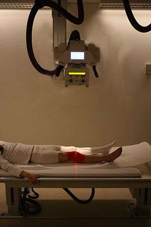
X-ray imaging

X-ray film
The first radiographs (X-ray images) were made by the action of X-rays on sensitized glass photographic plates. X-ray film (photographic film) soon replaced the glass plates, and film has been used for decades to acquire (and display) medical and industrial images. Gradually, digital computers gained the ability to store and display enough data to make digital imaging possible. Since the 1990s, computerized radiography and digital radiography have been replacing photographic film in medical and dental applications, though film technology remains in widespread use in industrial radiography processes (e.g. to inspect welded seams). The metal silver (formerly necessary to the radiographic & photographic industries) is a non-renewable resource although silver can easily be reclaimed from spent X-ray film. Where X-ray films required wet processing facilities, these new technologies do not. The digital archiving of images utilizing these new technologies also saves storage space. Thus it is beneficial that film use is being phased out.
Because photographic plates are sensitive to X-rays, they provide a means of recording the image, but they also require much X-ray exposure (to the patient). The addition of a fluorescent intensifying screen (or screens) in close contact with the film allows a lower dose to the patient, because the screen(s) improve the efficiency of x-ray detection, making more activation of the film from the same amount of x-rays, or the same activation of the film from a smaller amount of x-rays.

The part of the patient to be X-rayed is placed between the X-ray source and the image receptor to produce a shadow of the internal structure of that particular part of the body. X-rays are partially blocked ("attenuated") by dense tissues such as bone, and pass more easily through soft tissues. Areas where the X-rays strike darken when developed, causing bones to appear lighter than the surrounding soft tissue.
Contrast compounds containing barium or iodine, which are radiopaque, can be ingested in the gastrointestinal tract (barium) or injected in the artery or veins to highlight these vessels. The contrast compounds have high atomic numbered elements in them that (like bone) essentially block the X-rays and hence the once hollow organ or vessel can be more readily seen. In the pursuit of nontoxic contrast materials, many types of high atomic number elements were evaluated. Unfortunately, some elements chosen proved to be harmful – for example, thorium was once used as a contrast medium (Thorotrast) – which turned out to be toxic, causing a very high incidence of cancer decades after use. Modern contrast material has improved and, while there is no way to determine who may have a sensitivity to the contrast, the incidence of allergic reactions is low.
Photostimulable phosphors
An alternative method of recording the X-rays is the use of photostimulated luminescence (PSL), pioneered by Fuji in the 1980s. In modern hospitals a photostimulable phosphor plate (PSP plate) is used in place of the photographic plate. After the plate is X-rayed, excited electrons in the phosphor material remain 'trapped' in 'colour centres' in the crystal lattice until stimulated by a laser beam passed over the plate surface. The light given off during laser stimulation is collected by a photomultiplier tube, and the resulting signal is converted into a digital image by computer technology, which gives this process its common name, computed radiography. The PSP plate can be reused, and existing X-ray equipment requires no modification to use them.
Image intensifiers

X-rays are also used in "real-time" procedures such as angiography or contrast studies of the hollow organs (e.g. barium enema of the small or large intestine) using fluoroscopy acquired using an X-ray image intensifier. Angioplasty, medical interventions of the arterial system, rely heavily on X-ray-sensitive contrast to identify potentially treatable lesions.
Direct semiconductor detectors
Since the 1970s, new semiconductor detectors have been developed (silicon or germanium doped with lithium: Si(Li) or Ge(Li)). X-ray photons are converted to electron-hole pairs in the semiconductor and are collected to detect the X-rays. When the temperature is low enough (the detector is cooled by Peltier effect or even cooler liquid nitrogen), it is possible to directly determine the X-ray energy spectrum; this method is called energy dispersive X-ray spectroscopy (EDX or EDS); it is often used in small X-ray fluorescence spectrometers. These detectors are sometimes called "solid state detectors". Detectors based on cadmium telluride (CdTe) and its alloy with zinc, cadmium zinc telluride, have an increased sensitivity, which allows lower doses of X-rays to be used.
Practical application in medical imaging started in the 1990s. Currently amorphous selenium is used in commercial large area flat panel X-ray detectors for mammography and chest radiography. Current research and development is focused around energy resolving pixel detectors, such as CERN's Medipix detector and Science and Technology Facilities Council's HEXITEC detector.
A common semiconductor diode, such as a 1N4001, will produce a small amount of current when placed in an X-ray beam. A test device once used by medical imaging service personnel was a small project box that contained several diodes of this type connected in series, which could be connected to an oscilloscope as a quick diagnostic.
Silicon drift detectors (SDDs), produced by conventional semiconductor fabrication, now provide a cost-effective and high resolving power radiation measurement. Unlike conventional X-ray detectors, such as Si(Li), they do not need to be cooled with liquid nitrogen.
Scintillators
Some materials such as sodium iodide (NaI) can "convert" an X-ray photon to a visible photon; an electronic detector can be built by adding a photomultiplier. These detectors are called "scintillators", filmscreens, or "scintillation counters". The main advantage of using these is that an adequate image can be obtained while subjecting the patient to a much lower dose of X-rays.
In order to gain energy spectrum information, a diffracting crystal may be used to separate the different photons. The method is called wavelength dispersive X-ray spectroscopy (WDX or WDS). Position-sensitive detectors are often used in conjunction with dispersive elements. Other detection equipment that is inherently energy-resolving may be used, such as the aforementioned proportional counters. In either case, use of suitable pulse-processing (MCA) equipment allows digital spectra to be created for later analysis.
Scintillator plus semiconductor detectors
With the advent of large semiconductor array detectors it has become possible to design detector systems using a scintillator screen to convert from X-rays to visible light which is then converted to electrical signals in an array detector. Indirect flat panel detectors (FPDs) are in widespread use today in medical, dental, veterinary, and industrial applications.
The array technology is a variant on the amorphous silicon TFT arrays used in many flat panel displays, like the ones in computer laptops. The array consists of a sheet of glass covered with a thin layer of silicon that is in an amorphous or disordered state. At a microscopic scale, the silicon has been imprinted with millions of transistors arranged in a highly ordered array, like the grid on a sheet of graph paper. Each of these thin-film transistors (TFTs) is attached to a light-absorbing photodiode making up an individual pixel (picture element). Photons striking the photodiode are converted into two carriers of electrical charge, called electron-hole pairs. Since the number of charge carriers produced will vary with the intensity of incoming light photons, an electrical pattern is created that can be swiftly converted to a voltage and then a digital signal, which is interpreted by a computer to produce a digital image. Although silicon has outstanding electronic properties, it is not a particularly good absorber of X-ray photons. For this reason, X-rays first impinge upon scintillators made from such materials as gadolinium oxysulfide or caesium iodide. The scintillator absorbs the X-rays and converts them into visible light photons that then pass onto the photodiode array.
Dose measurement
Gas detectors

X-rays going through a gas will ionize it, producing positive ions and free electrons. An incoming photon will create a number of such ion pairs proportional to its energy. If there is an electric field in the gas chamber ions and electrons will move in different directions and thereby cause a detectable current. The behaviour of the gas will depend on the applied voltage and the geometry of the chamber. This gives rise to a few different types of gas detectors described below.
Ionization chambers use a relatively low electric field of about 100 V/cm to extract all ions and electrons before they recombine.[1] This gives a steady current proportional to the dose rate the gas is exposed to. Ion chambers are widely used as hand held radiation survey meters to check radiation dose levels.
Proportional counters use a geometry with a thin positively charged anode wire in the center of a cylindrical chamber. Most of the gas volume will act as an ionization chamber, but in the region closest to the wire the electric field is high enough to make the electrons ionize gas molecules. This will create an avalanche effect greatly increasing the output signal. Since every electron cause an avalanche of approximately the same size the collected charge is proportional to the number of ion pairs created by the absorbed x-ray. This makes it possible to measure the energy of each incoming photon.
Geiger–Müller counters use an even higher electric field so that UV-photons are created. These start new avalanches, eventually resulting in a total ionization of the gas around the anode wire. This makes the signal very strong, but causes a dead time after each event and makes it impossible to measure the X-ray energies.
Gas detectors are usually single pixel detectors measuring only the average dose rate over the gas volume or the number of interacting photons as explained above, but they can be made spatially resolving by having many crossed wires in a wire chamber.
Silicon PN solar cells
It was demonstrated in the 1960s that silicon PN solar cells are suitable for detection of all forms of ionizing radiation including extreme UV, soft X-rays, and hard X-rays. This form of detection operates via photoionization, a process where ionizing radiation strikes an atom and releases a free electron.[2] This type of broadband ionizing radiation sensor requires a solar cell, an ammeter, and a visible light filter on top of the solar cell that allows the ionizing radiation to hit the solar cell while blocking unwanted wavelengths.
References
- ↑ Albert C. Thompson. X-Ray Data Booklet, Section 4-5: X-ray detectors (PDF).
- ↑ Photovoltaic Effect Produced in Silicon Solar Cells by x-ray and Gamma-Rays, Karl Scharf, January 25, 1960, Journal of Research of the National Bureau of Standards