Biotin
 | |
 | |
| Names | |
|---|---|
| IUPAC name
5-[(3aS,4S,6aR)-2-oxohexahydro-1H-thieno[3,4-d]imidazol-4-yl]pentanoic acid | |
| Other names
Vitamin B7; Vitamin H; Coenzyme R; Biopeiderm | |
| Identifiers | |
| 58-85-5 | |
| 3D model (Jmol) | Interactive image Interactive image |
| ChEBI | CHEBI:15956 |
| ChEMBL | ChEMBL857 |
| ChemSpider | 149962 |
| DrugBank | DB00121 |
| ECHA InfoCard | 100.000.363 |
| 4787 | |
| KEGG | D00029 |
| PubChem | 171548 |
| UNII | 6SO6U10H04 |
| |
| |
| Properties | |
| C10H16N2O3S | |
| Molar mass | 244.31 g·mol−1 |
| Appearance | White crystalline needles |
| Melting point | 232 to 233 °C (450 to 451 °F; 505 to 506 K) |
| 22 mg/100 mL | |
| Pharmacology | |
| A11HA05 (WHO) | |
| Hazards | |
| NFPA 704 | |
| Except where otherwise noted, data are given for materials in their standard state (at 25 °C [77 °F], 100 kPa). | |
| | |
| Infobox references | |

Biotin is a water-soluble B-vitamin, also called vitamin B7 and formerly known as vitamin H or coenzyme R.[2]
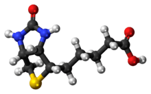
It is composed of a ureido ring fused with a tetrahydrothiophene ring. A valeric acid substituent is attached to one of the carbon atoms of the tetrahydrothiophene ring. Biotin is a coenzyme for carboxylase enzymes, involved in the synthesis of fatty acids, isoleucine, and valine, and in gluconeogenesis.
Biotin deficiency can be caused by inadequate dietary intake or inheritance of one or more inborn genetic disorders that affect biotin metabolism. Subclinical deficiency can cause mild symptoms, such as hair thinning or skin rash typically on the face. Neonatal screening for biotinidase deficiency began in the United States in 1984 and today many countries test for this disorder at birth. Individuals born prior to 1984 are unlikely to have been screened, thus the true prevalence of the disorder is unknown.
General

Biotin is necessary for cell growth, the production of fatty acids, and the metabolism of fats and amino acids.[2] Biotin assists in various metabolic reactions involving the transfer of carbon dioxide. It may also be helpful in maintaining a steady blood sugar level. Biotin is often recommended as a dietary supplement for strengthening hair and nails, though scientific data supporting this outcome are weak.[2] Nevertheless, biotin is found in many cosmetics and health products for the hair and skin.[3][4]
Biotin deficiency is rare. The amounts needed are small, a wide range of foods contain biotin, and intestinal bacteria synthesize biotin, which is then absorbed by the host animal. For that reason, statutory agencies in many countries, for example the USA[5] and Australia,[6] have not formally established a recommended daily intake of biotin. Instead, an Adequate Intake (AI) is identified based on the theory that average intake meets needs. Future research could result in biotin AIs with EARs and RDAs (see Dietary Reference Intake section).
A number of rare metabolic disorders exist in which an individual's metabolism of biotin is abnormal, such as deficiency in the holocarboxylase synthetase enzyme which covalently links biotin onto the carboxylase, where the biotin acts as a cofactor.[7]
Biosynthesis
Biotin has an unusual structure (see above figure), with two rings fused together via one of their sides. The two rings are ureido and thiophene moieties. Biotin is a heterocyclic, S-containing monocarboxylic acid. It is made from two precursors, alanine and pimeloyl-CoA via three enzymes. 8-Amino-7-oxopelargonic acid synthase is a pyridoxal 5'-phosphate enzyme. The pimeloyl-CoA, could be produced by a modified fatty acid pathway involving a malonyl thioester as the starter. 7,8Diaminopelargonic acid (DAPA) aminotransferase is unusual in using S-adenosyl methionine (SAM) as the NH2 donor. Dethiobiotin synthetase catalyzes the formation of the ureido ring via a DAPA carbamate activated with ATP. Biotin synthase reductively cleaves SAM into a deoxyadenosyl radical—a first radical formed on dethiobiotin is trapped by the sulfur donor, which was found to be the iron-sulfur (Fe-S) center contained in the enzyme.[8]
Cofactor biochemistry
D-(+)-Biotin is a cofactor responsible for carbon dioxide transfer in several carboxylase enzymes:
- Acetyl-CoA carboxylase alpha
- Acetyl-CoA carboxylase beta
- Methylcrotonyl-CoA carboxylase
- Propionyl-CoA carboxylase
- Pyruvate carboxylase
Biotin is important in fatty acid synthesis, branched-chain amino acid catabolism, and gluconeogenesis.[2] It covalently attaches to the epsilon-amino group of specific lysine residues in these carboxylases. This biotinylation reaction requires ATP and is catalyzed by holocarboxylase synthetase.[9] In bacteria, biotin is attached to biotin carboxyl carrier protein (BCCP) by biotin protein ligase (BirA in E. coli).[10] The attachment of biotin to various chemical sites, biotinylation, is used as an important laboratory technique to study various processes, including protein localization, protein interactions, DNA transcription, and replication. Biotinidase itself is known to be able to biotinylate histone proteins,[11] but little biotin is found naturally attached to chromatin.
Biotin binds tightly to the tetrameric protein avidin (also streptavidin and neutravidin), with a dissociation constant Kd on the order of 10−15 M, which is one of the strongest known protein-ligand interactions.[12] This is often used in different biotechnological applications. Until 2005, very harsh conditions were thought to be required to break the biotin-streptavidin bond.[13]
Dietary Reference Intake
The Food and Nutrition Board of the U.S. Institute of Medicine updated Estimated Average Requirements (EARs) and Recommended Dietary Allowances (RDAs) for B vitamins in 1998. At that time there was not sufficient information to establish EARs and RDAs for biotin. In instances such as this, the Board sets Adequate Intakes (AIs), with the understanding that at some later date, AIs will be replaced by more exact information. The current AI for adults ages 19 and up is 30 μg/day. AI for pregnancy is 30 μg/day. AI for lactation is 35 μg/day. For infants up to 12 months the AI is 5-6 μg/day For children ages 1–18 years the AI increases with age from 8 to 25 μg/day.[14]
As for safety, the FNB sets Tolerable Upper Intake Levels (known as ULs) for vitamins and minerals when evidence is sufficient. In the case of biotin there is no UL, as there is insufficient human data to identify adverse effects from high doses. The European Food Safety Authority reviewed the same safety question and also reached the conclusion that there was not sufficient evidence to set a UL for biotin.[15] Collectively the EARs, RDAs, AIs and ULs are referred to as Dietary Reference Intakes.
For U.S. food and dietary supplement labeling purposes the amount in a serving is expressed as a percent of Daily Value (%DV). For biotin labeling purposes 100% of the Daily Value was 300 μg, but as of May 2016 it has been revised to 30 μg to bring it into agreement with the AI. A table of the pre-change adult Daily Values is provided at Reference Daily Intake. Food and supplement companies have until July 2018 to comply with the change.
Sources
Biotin is consumed from a wide range of food sources in the diet, but few are particularly rich sources. Foods with a relatively high biotin content include peanuts, Swiss chard and other leafy green vegetables, raw egg yolk (however, the consumption of avidin-containing egg whites with egg yolks minimizes the effectiveness of egg yolk's biotin in one's body), liver, and Saskatoon berries. The dietary biotin intake in Western populations has been estimated to be 35 to 70 μg/d (143–287 nmol/d).[16]
Biotin is also available in supplement form and can be found in most pharmacies. The synthetic process developed by Leo Sternbach and Moses Wolf Goldberg in the 1940s uses fumaric acid as a starting material.[17]
Import into the cell
In mammals biotin is imported into cells by the Na+-dependent multivitamin (pantothenate, biotin, lipoate) transporter (SMVT).[18]
In bacteria, several families of proteins, especially the BioY family of transporters, imports biotin into cells.[19]
Bioavailability
Biotin is also called vitamin H (the H represents Haar und Haut, German words for "hair and skin") or vitamin B7. Studies on its bioavailability have been conducted in rats and in chicks. Based on these studies, biotin bioavailability may be low or variable, depending on the type of food being consumed. In general, biotin exists in food as protein-bound form or biocytin.[20] Proteolysis by protease is required prior to absorption. This process assists free biotin release from biocytin and protein-bound biotin. The biotin present in corn is readily available; however, most grains have about a 20-40% bioavailability of biotin.[21]
The wide variability in biotin bioavailability may be due to the ability of an organism to break various biotin-protein bonds from food. Whether an organism has an enzyme with that ability will determine the bioavailability of biotin from the foodstuff.[21]
Factors that affect biotin requirements
The frequency of marginal biotin status is not known, but the incidence of low circulating biotin levels in alcoholics has been found to be much greater than in the general population. Also, relatively low levels of biotin have been reported in the urine or plasma of patients who have had a partial gastrectomy or have other causes of achlorhydria, burn patients, epileptics, elderly individuals, and athletes.[21] Pregnancy and lactation may be associated with an increased demand for biotin. In pregnancy, this may be due to a possible acceleration of biotin catabolism, whereas, in lactation, the higher demand has yet to be elucidated. Recent studies have shown marginal biotin deficiency can be present in human gestation, as evidenced by increased urinary excretion of 3-hydroxyisovaleric acid, decreased urinary excretion of biotin and bisnorbiotin, and decreased plasma concentration of biotin. Additionally, smoking may further accelerate biotin catabolism in women.[22]
Deficiency
Biotin deficiency typically occurs from dietary absence of the vitamin. Consuming raw egg whites over months may result in biotin deficiency.[2]
Deficiency can be addressed with nutritional supplementation.[2]
Deficiency symptoms include:
- Brittle and thin fingernails
- Hair loss (alopecia)
- Conjunctivitis
- Dermatitis in the form of a scaly, red rash around the eyes, nose, mouth, and genital area.
- Neurological symptoms in adults, such as depression, lethargy, hallucination, and numbness and tingling of the extremities[2]
The neurological and psychological symptoms can occur with only mild deficiencies. Dermatitis, conjunctivitis, and hair loss will generally occur only when deficiency becomes more severe.[2]
Individuals with hereditary disorders of biotin deficiency have evidence of impaired immune system function, including increased susceptibility to bacterial and fungal infections.[23]
Pregnant women tend to have a high risk of biotin deficiency. Nearly half of pregnant women have abnormal increases of 3-hydroxyisovaleric acid, which reflects reduced status of biotin.[23] Several studies have reported this possible biotin deficiency during the pregnancy may cause infants' congenital malformations, such as cleft palate. Mice fed with dried raw egg to induce biotin deficiency during the gestation resulted in up to 100% incidence of the infants' malnourishment. Infants and embryos are more sensitive to the biotin deficiency. Therefore, even a mild level of the mother's biotin deficiency that does not reach the appearance of physiological deficiency signs may cause a serious consequence in the infants.
Metabolic disorders
Inherited metabolic disorders characterized by deficient activities of biotin-dependent carboxylases are termed multiple carboxylase deficiency. These include deficiencies in the enzymes holocarboxylase synthetase or biotinidase. Holocarboxylase synthetase deficiency prevents the body's cells from using biotin effectively, and thus interferes with multiple carboxylase reactions.[24] Biochemical and clinical manifestations include: ketolactic acidosis, organic aciduria, hyperammonemia, skin rash, feeding problems, hypotonia, seizures, developmental delay, alopecia, and coma.
Biotinidase deficiency is not due to inadequate biotin, but rather to a deficiency in the enzymes that process it. Biotinidase catalyzes the cleavage of biotin from biocytin and biotinyl-peptides (the proteolytic degradation products of each holocarboxylase) and thereby recycles biotin. It is also important in freeing biotin from dietary protein-bound biotin.[24] General symptoms include decreased appetite and growth. Dermatologic symptoms include dermatitis, alopecia, and achromotrichia (absence or loss of pigment in the hair).[25] Perosis (a shortening and thickening of bones) is seen in the skeleton. Fatty liver and kidney syndrome and hepatic steatosis also can occur.[21]
Use in biotechnology
Biotin is widely used throughout the biotechnology industry to conjugate proteins for biochemical assays.[26] Biotin's small size means the biological activity of the protein will most likely be unaffected. This process is called biotinylation. Because both streptavidin and avidin bind biotin with high affinity (Kd of 10−14 mol/l to 10−15 mol/l) and specificity, biotinylated proteins of interest can be isolated from a sample by exploiting this highly stable interaction. The sample is incubated with streptavidin/avidin beads, allowing capture of the biotinylated protein of interest. Any other proteins binding to the biotinylated molecule will also stay with the bead and all other unbound proteins can be washed away. However, due to the extremely strong streptavidin-biotin interaction, very harsh conditions are needed to elute the biotinylated protein from the beads (typically 6M guanidine HCl at pH 1.5), which often will denature the protein of interest. To circumvent this problem, beads conjugated to monomeric avidin can be used, which has a decreased biotin-binding affinity of ~10−8 mol/l, allowing the biotinylated protein of interest to be eluted with excess free biotin.
As one of the strongest non-covalent interactions, the binding of biotin to streptavidin is commonly used as the target molecular interaction in the research of biosensors and cell sorting.[27][28]
ELISAs often make use of biotinylated secondary antibodies against the antigen of interest, followed by a detection step using streptavidin conjugated to a reporter molecule, such as horseradish peroxidase or alkaline phosphatase.
Toxicity
Animal studies have indicated few, if any, effects due to high level doses of biotin. This may provide evidence that both animals and humans could tolerate doses of at least an order of magnitude greater than each of their nutritional requirements. There are no reported cases of adverse effects from receiving high doses of the vitamin, in particular, when used in the treatment of metabolic disorders causing sebhorrheic dermatitis in infants.[29] However, excess biotin accumulation can inhibit endogenous sirtuin activity leading to increased inflammation, cellularity, and collagen deposition, and may be partly responsible for age related metabolic problems. Reversed by calorie restriction in mice.[30]
See also
References
- ↑ Merck Index, 11th Edition, 1244.
- 1 2 3 4 5 6 7 8 "Biotin: MedlinePlus Supplements". 13 September 2013. Retrieved 2013-09-29.
- ↑ . PMID 11800048. Missing or empty
|title=(help) - ↑ "Vitamin H (Biotin)". University of Maryland Medical Center. 1 June 2011. Retrieved 4 May 2012.
- ↑ Otten, JJ; Hellwig, JP; Meyers, LD., eds. (2006). Dietary Reference Intakes: The Essential Guide to Nutrient Requirements. The National Academies Press. ISBN 0-309-10091-7.
- ↑ National Health and Medical Research Council: Nutrient Reference Values for Australia and New Zealand
- ↑ Zempleni J, Hassan YI, Wijeratne SS (2008). "Biotin and biotinidase deficiency". Expert Rev Endocrinol Metab. 3 (6): 715–724. doi:10.1586/17446651.3.6.715. PMC 2726758
 . PMID 19727438.
 - ↑ Marquet A, Bui BT, Florentin D (2001). "Biosynthesis of biotin and lipoic acid". Vitam. Horm. Vitamins & Hormones. 61: 51–101. doi:10.1016/S0083-6729(01)61002-1. ISBN 978-0-12-709861-6. PMID 11153271.
- ↑ Zempleni J, Wijeratne SS, Hassan YI (2009). "Biotin". BioFactors. 35 (1): 36–46. doi:10.1002/biof.8. PMID 19319844.
- ↑ Chapman-Smith A, Cronan JE (1999). "Molecular biology of biotin attachment to proteins". J. Nutr. 129 (2S Suppl): 477S–484S. PMID 10064313.
- ↑ Hymes J, Fleischhauer K, Wolf B (1995). "Biotinylation of histones by human serum biotinidase: assessment of biotinyl-transferase activity in sera from normal individuals and children with biotinidase deficiency". Biochem Mol Med. 56 (1): 76–83. doi:10.1006/bmme.1995.1059. PMID 8593541.
- ↑ Laitinen OH, Hytönen VP, Nordlund HR, Kulomaa MS (2006). "Genetically engineered avidins and streptavidins". Cell Mol Life Sci. 63 (24): 2992–3017. doi:10.1007/s00018-006-6288-z. PMID 17086379.
- ↑ Holmberg A, Blomstergren A, Nord O, Lukacs M, Lundeberg J, Uhlén M (2005). "The biotin-streptavidin interaction can be reversibly broken using water at elevated temperatures". Electrophoresis. 26 (3): 501–10. doi:10.1002/elps.200410070. PMID 15690449.
- ↑ "Biotin". In: Dietary Reference Intakes for Thiamin, Riboflavin, Niacin, Vitamin B6, Folate, Vitamin B12, Pantothenic Acid, Biotin, and Choline. National Academy Press. 1998. pp. 374-389.
- ↑ Tolerable Upper Intake Levels For Vitamins And Minerals (PDF), European Food Safety Authority, 2006
- ↑ Zempleni J, Mock DM (1999). "Biotin biochemistry and human requirements". J Nutr Biochem. 10 (3): 128–138. doi:10.1016/S0955-2863(98)00095-3. PMID 15539280.
- ↑ "Biotin". HQhair.org. 2014-03-19. Retrieved 2014-03-19.
- ↑ Na+-dependent multivitamin (pantothenate, biotin, lipoate) transporter in the Transporter Classification Database, accessed 7 Jan 2016
- ↑ biotin transporters in the Transporter Classification Database, accessed 7 Jan 2016
- ↑ Gropper SS, Smith JL, Groff JL (2005). Advanced nutrition and human metabolism. Belmont. ISBN 0-534-55986-7.
- 1 2 3 4 Combs, Gerald F. Jr. (2008). The Vitamins: Fundamental Aspects in Nutrition and Health. San Diego: Elsevier, Inc. ISBN 978-0-12-183493-7.
- ↑ Bowman, BA; Russell, RM., eds. (2006). "Biotin". Present Knowledge in Nutrition, Ninth Edition, Vol 1. Washington, DC: International Life Sciences Institute. ISBN 978-1-57881-198-4.
- 1 2 Higdon, Jane (2016). "Biotin". Linus Pauling Institute, Micronutrient Information Center, Oregon State University, Corvallis. Retrieved 12 October 2016.
- 1 2 Wolf B, Grier RE, Secor McVoy JR, Heard GS (1985). "Biotinidase deficiency: a novel vitamin recycling defect". J Inherit Metab Dis. 8 (1): 53–8. doi:10.1007/BF01800660. PMID 3930841.
- ↑ biology-online.org
- ↑ "Overview of Protein Labeling". Thermo Fisher Scientific. Retrieved 22 April 2012.
- ↑ Xu, Zhida; Xinhao, Wang; Han, Kevin; Li, Shuo; Liu, Logan (2013). "Elastomeric 2D grating and hemispherical optofluidic chamber for multifunctional fluidic sensing". Journal of the Optical Society of America A. 30 (12). pp. 2466–2472. doi:10.1364/JOSAA.30.002466.
- ↑ Xu, Zhida; Jiang, Jing; Wang, Xinhao; Han, Kevin; Ameen, Abid; Khan, Ibrahim; Chang, Te-Wei; Liu, Logan (2016). "Large-area, uniform and low-cost dual-mode plasmonic naked-eye colorimetry and SERS sensor with handheld Raman spectrometer". Nanoscale. 8 (11): 6162–6172. doi:10.1039/C5NR08357E.
- ↑ Combs, Gerald F. Jr. (1998). The Vitamins: Fundamental Aspects in Nutrition and Health. Ithaca: Elsevier Academic Press. ISBN 0-12-183492-1.pg. 360
- ↑ Diabetes (2015);64:1-15