Vastus medialis
| Vastus medialis | |
|---|---|
 Muscles of lower extremity | |
| Details | |
| Origin | Medial side of femur |
| Insertion | Quadriceps tendon |
| Artery | Femoral artery |
| Nerve | Femoral nerve |
| Actions | Extends leg |
| Identifiers | |
| Latin | Musculus vastus medialis or musculus vastus internus |
| TA | A04.7.02.023 |
| FMA | 22432 |
The vastus medialis (/ˈvæstəs ˌmɛdiˈeɪlᵻs/ or /ˈvæstəs ˌmɛdiˈælᵻs/) (vastus internus or teardrop muscle) is an extensor muscle located medially in the thigh that extends the knee. The vastus medialis is part of the quadriceps muscle group.
Structure
- The vastus medialis is a muscle present in the anterior compartment of thigh, and is one of the four muscles that make up the quadriceps muscle. The others being the vastus lateralis, vastus intermedius and rectus femoris.[1] It is the most medial of the "vastus" group of muscles. The vastus medialis arises medially along the entire length of the femur, and attaches with the other muscles of the quadriceps in the quadriceps tendon.[1]
- The vastus medialis muscle originates from a continuous line of attachment on the femur, which begins on the front and middle side (anteromedially) on the intertrochanteric line of the femur. It continues down and back (posteroinferiorly) along the pectineal line and then descends along the inner (medial) lip of the linea aspera and onto the medial supracondylar line of the femur. The fibers converge onto the inner (medial) part of the quadriceps tendon and the inner (medial) border of the patella.[1]
- The obliquus genus muscle is the most distal segment of the vastus medialis muscle. Its specific training plays an important role in maintaining patella position and limiting injuries to the knee. With no clear delineation, it is simply the most distal group of fibers of the vastus medialis.
Function
The vastus medialis is one of five muscles in the anterior compartment of the thigh.[1] It is involved in knee extension, along with the other muscles which make up the quadriceps muscle.[1] The vastus medialis also contributes to correct tracking of the patella.[2]
A division of the vastus medialis muscle into two groups of fibers has been hypothesized, a long and relatively inline group of fibres with the quadriceps ligament, the vastus medialis longus; and a shorter and more obliquely oriented with group of fibbres, the vastus medialis obliquus. There is as yet insufficient evidence to conclusively confirm or deny this hypothesis.[3]
Clinical significance
Knee pain
Knee pain is thought to be primarily associated with specific quadriceps muscle weakness or fatigue, especially in the vastus medialis obliquus (VMO). It is known that fatigue can be caused by many different mechanisms, ranging from the accumulation of metabolites within muscle fibers to the generation of an inadequate motor command in the motor cortex.[4] Characteristics of the vastus medialis, including its angle of insertion, correlate with presence of knee joint pain (patellofemoral pain syndrome).[5] However, this syndrome is complex and definitive evidence of causality has not yet been published.
Misfiring and fatiguing of the VMO causes mal-tracking of the patella and subsequent damage to surrounding structures creating increased force on the knees, often resulting in injuries such as patellofemoral pain syndrome, anterior cruciate ligament rupture, chondromalacia, and tendinitis.[6] Through the use of electromyography, researchers can evaluate and record the electrical activity produced by the skeletal muscle of the VMO to analyze the biomechanics and detect any possible abnormalities, weakness, or fatigue. With an analysis of muscle activity of the VMO through the use of electromyography, proper rehabilitative plans and goals can be established to not only correct the already established abnormality, but even prevent such injuries if tested sooner. Preventing injuries is crucial as well as teaching proper training techniques to ensure there are no valgus collapse forces causing unplanned stress on other structures of the knee, causing asymmetry, and predisposing that individual for injury.
Additional images
 Right femur. Posterior surface.
 Muscles of the iliac and anterior femoral regions.
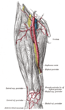 The femoral artery.
 Vastus medialis
 Vastus medialis muscle
 Vastus medialis muscle
 Vastus medialis muscle
 Vastus medialis muscle
 Vastus medialis muscle
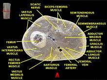 Muscles of thigh. Cross section.
See also
References
This article incorporates text in the public domain from the 20th edition of Gray's Anatomy (1918)
- 1 2 3 4 5 Drake, Richard L.; Vogl, Wayne; Tibbitts, Adam W.M. Mitchell; illustrations by Richard; Richardson, Paul (2005). Gray's anatomy for students. Philadelphia: Elsevier/Churchill Livingstone. pp. 518–519. ISBN 978-0-8089-2306-0.
- ↑ Sheehan, FT; Borotikar, BS; Behnam, AJ; Alter, KE (2012). "Alterations in in vivo knee joint kinematics following a femoral nerve branch block of the vastus medialis: Implications for patellofemoral pain syndrome". Clinical biomechanics. 27 (6): 525–31. doi:10.1016/j.clinbiomech.2011.12.012. PMC 3328589
 . PMID 22244738.
 - ↑ Smith, TO; Nichols, R; Harle, D; Donell, ST (2009). "Do the vastus medialis obliquus and vastus medialis longus really exist? A systematic review". Clinical Anatomy. 22 (2): 183–99. doi:10.1002/ca.20737. PMID 19090000.
- ↑ Enoka, RM; Duchateau, J (2008). "Muscle fatigue: what, why, and how it influences muscle function". The Journal of Physiology. 586 (1): 11–23. doi:10.1113/jphysiol.2007.139477. PMC 2375565. PMID 17702815.
- ↑ Jan, MH; Lin, DH; Lin, JJ; Lin, CH; Cheng, CK; Lin, YF (2009). "Differences in sonographic characteristics of the vastus medialis obliquus between patients with patellofemoral pain syndrome and healthy adults". The American journal of sports medicine. 37 (9): 1743–9. doi:10.1177/0363546509333483. PMID 19521000.
- ↑ Lefebvre, R; Leroux, A; Poumarat, G; Galtier, B; Guillot, M; Vanneuville, G; Boucher, JP (2006). "Vastus medialis: anatomical and functional considerations and implications based upon human and cadaveric studies". Journal of Manipulative and Physiological Therapeutics. 29 (2): 139–144. doi:10.1016/j.jmpt.2005.12.006. PMID 16461173.
External links
| Wikimedia Commons has media related to Vastus medialis. |
- The KNEEguru - educational site packed with knee content with sections on the quadriceps muscle
- Cross section image: pembody/body18b - Plastination Laboratory at the Medical University of Vienna
- PTCentral