Trichobilharzia regenti
| Trichobilharzia regenti | |
|---|---|
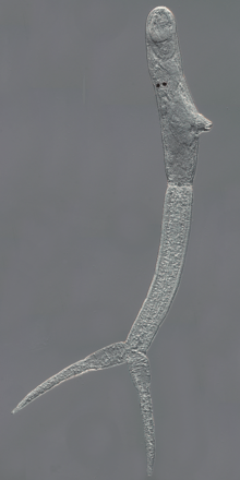 | |
| Trichobilharzia regenti, cercaria | |
| Scientific classification | |
| Kingdom: | Animalia |
| Phylum: | Platyhelminthes |
| Class: | Trematoda |
| Order: | Strigeidida |
| Family: | Schistosomatidae |
| Genus: | Trichobilharzia |
| Species: | T. regenti |
| Binomial name | |
| Trichobilharzia regenti Horák, Kolářová & Dvořák, 1998 [1] | |
Trichobilharzia regenti is a neuropathogenic parasitic flatworm of birds which also causes cercarial dermatitis in humans.[2] The species was originally described in 1998 in the Czech Republic[1] and afterwards it was detected also in other European countries, e.g. Denmark,[3] France,[4] Iceland[5] or Russia,[6] and even in Iran.[7] For its unique neurotropic behaviour in vertebrate hosts, the host-parasite interactions are extensively studied in terms of molecular biology, biochemistry and immunology.[8][9]
Life cycle
The life cycle of T. regenti is analogous to that of human schistosomes. Adult flukes mate in a nasal mucosa of anatid birds (e.g. Anas platyrhynchos and Cairina moschata) and produce eggs with miracidia which hatch directly in the host tissue and leak outside when the bird is drinking/feeding.[1] Once in water, the miracidia swim using their cilia and actively search for a proper molluscan intermediate host (Radix lagotis, Radix labiata, Radix peregra).[10] In the snail, the miracidia develop into a primary sporocyst in which secondary sporocysts are formed and give rise to cercariae later on.[11]
Cercariae, infective larvae, exit the snail and penetrate the skin of an avian host. After penetration, they transform to schistosomula (subadult stage) and look for peripheral nerves to use them to get to the spinal cord. Through it they continue their migration to the brain[12][13] and, finally, the nasal tissue in a bill. Here, they mature, copulate and lay eggs while causing pathology (inflammatory infiltration, haemorrhages).[14]
If mammals are infected by cercariae (instead of birds), the parasites die in the skin being entrapped by immune response.[15] The clinical manifestation of such infection is known as an neglected allergic disease called cercarial dermatitis (or swimmer's itch).[16] In mice, especially in immunodeficient ones, migration of the parasite to the spinal cord was observed.[17][18]
Migration in vertebrate hosts
When cercariae of T. regenti find either avian or mammalian host, they penetrate its skin. For this purpose, they are equipped with cysteine peptidases present in their excretory/secretory products, which are capable of keratin and collagen degradation.[19][20] Experiments with laboratory prepared recombinant form of the cysteine peptidase cathepsin B2 of T. regenti (TrCB2) confirmed its ability to cleave skin proteins (collagen, keratin and elastin).[21]
After penetration the skin, cercariae transform to schistosomula and start a migration through the host’s body. They avoid penetration into blood capillaries and rather prefer entering peripheral nerves in host’s limbs. Schistosomula are found in peripheral nerves of ducks and mice as soon as 1.5 and 1 day post infection (DPI), respectively.[13] In both types of hosts, schistosomula exhibit a high affinity to the central nervous system which they enter via spinal roots.[22] Based on recent observation by 3D imaging techniques (ultramicroscopy and micro-CT), schistosomula appear to migrate preferably through the white matter of the spinal cord in both birds and mammals.[23]
The next course of the infection differs in final and accidental hosts. In ducks, schistosomula are observed in synsacral segments of a spinal cord 3 DPI and 7–8 days latter (10–11 DPI) they reach the brain. In their final localisation (the nasal tissue), they occur 13–14 DPI and laying eggs starts 15 DPI.[13][14] In mice, the first schistosomula are found in a lumbar spinal cord as early as 2 DPI and medulla oblongata is invaded the day after, but only in some individuals. Most of schistosomula stay localised in the thoracic and cervical spinal cord and only exceptionally migrate to the brain.[12][13] Neither the presence of worms has been detected in a nasal cavity nor has their maturation been noticed in the nervous tissue. Schistosomula development in mice is suppressed likely due to the host immune response and/or the presence/absence of some essential (nutritional, stimulatory) host factors.[18]
Pathology in vertebrate hosts

In vertebrate hosts infected by T. regenti, pathological states might be caused by:
- penetrating cercariae transforming to schistosomula in the skin,
- schistosomula migrating through the central nervous system,
- adults laying eggs in nasal mucosa (only in avian hosts).

Although mice are accidental hosts, most of the studies dealing with the pathological effects of T. regenti were conducted on this model.
In the initial phase of the infection, early transformed schistosomula are localised in the skin. Information about the immune response in the skin of birds has not been completed yet. In mice, immediate oedema and thickening of the site appear as early as 30 minutes after the penetration of cercariae; erythema is evident as well. Within 48 hours, inflammatory foci containing neutrophils, eosinophils, macrophages, CD4+ lymphocytes and degranulating mast cells develop around the parasites.[15][17] In case of repeated infections, the cellular infiltration is substantially elevated and the extensive inflammation may lead to formation of large abscesses or even epidermal and/or dermal necrosis.[15] In humans, the clinical symptoms of cercarial penetration consist of macules/papules formation at the sites where the parasite entered the skin accompanied by intensive itching. The manifestation is more severe in previously sensitised people. This disease caused not only by T. regenti but also by cercariae of other bird schistosome species is called cercarial dermatitis. It is regarded as a neglected allergic disease.[16]

The next phase of T. regenti infection is represented by schistosomula migration in the central nervous system. This is accompanied by serious neurological malfunctions in birds that suffer from leg paralysis and balance disorders.[12] At this stage, schistosomula feed on nervous tissue as demonstrated by detection of oligodendrocytes and neurons in the lumen of parasite’s intestine.[22] A cysteine peptidase cathepsin B1 of T. regenti (TrCB1) localised in intestines of migrating schistosomula is capable of myelin basic protein degradation, thus probably serving for nervous tissue digestion.[24] Nonetheless, the nervous tissue ingestion has likely only a minor pathogenic effect on the host central nervous tissue.[22] This is underpinned by observations of leg paralysis only in immunocompromised hosts,[17][22] whereas in experiments with immunocompetent mouse strains, the infected animals did not reveal any neurological disorders.[13][17][22] The neurological symptoms originate probably in mechanical damage of the nervous tissue leading to dystrophic or even necrotic changes of neurons and axonal injury. The cause of it is large migrating schistosomula (approximately 340×80 μm) which are not destroyed by proper immune response.[17][22][25]
In avian hosts, T. regenti reaches the nasal tissue where it mates and lay eggs. The gross pathology at this site consists of focal haemorrhages dispersed all over the mucosa. Infiltrates of lymphocytes are present around the eggs and even granulomas containing lymphocytes, eosinophils and heterophils form at later phases. Similar infiltrates are present around free miracidia, but the granuloma formation was not recorded. Interestingly, no cell reaction was noted in the vicinity of adult worms.[14]
References
- 1 2 3 Horák, Petr; Kolářová, Libuše; Dvořák, Jan (1998-12-01). "Trichobilharzia regenti n. sp. (Schistosomatidae, Bilharziellinae), a new nasal schistosome from Europe". Parasite. 5 (4): 349–357. doi:10.1051/parasite/1998054349. ISSN 1252-607X. PMID 9879557.
- ↑ Horák, Petr; Mikeš, Libor; Lichtenbergová, Lucie; Skála, Vladimír; Soldánová, Miroslava; Brant, Sara Vanessa (2015-01-01). "Avian schistosomes and outbreaks of cercarial dermatitis". Clinical Microbiology Reviews. 28 (1): 165–190. doi:10.1128/CMR.00043-14. ISSN 1098-6618. PMC 4284296
 . PMID 25567226.
 - ↑ Christiansen, Anne Ø; Olsen, Annette; Buchmann, Kurt; Kania, Per W.; Nejsum, Peter; Vennervald, Birgitte J. (2016-03-01). "Molecular diversity of avian schistosomes in Danish freshwater snails". Parasitology Research. 115 (3): 1027–1037. doi:10.1007/s00436-015-4830-3. ISSN 1432-1955. PMID 26573519.
- ↑ Jouet, Damien; Skírnisson, Karl; Kolárová, Libuse; Ferté, Hubert (2010-09-01). "Final hosts and variability of Trichobilharzia regenti under natural conditions". Parasitology Research. 107 (4): 923–930. doi:10.1007/s00436-010-1953-4. ISSN 1432-1955. PMID 20556426.
- ↑ Skírnisson, Karl; Kolářová, Libuse; Horák, Petr; Ferté, Hubert; Jouet, Damien (2012-05-01). "Morphological features of the nasal blood fluke Trichobilharzia regenti (Schistosomatidae, Digenea) from naturally infected hosts". Parasitology Research. 110 (5): 1881–1892. doi:10.1007/s00436-011-2713-9. ISSN 1432-1955. PMID 22146993.
- ↑ Korsunenko, A. V.; Chrisanfova, G. G.; Ryskov, A. P.; Movsessian, S. O.; Vasilyev, V. A.; Semyenova, S. K. (2010-08-01). "Detection of European Trichobilharzia schistosomes ( T. franki , T. szidati , and T. regenti ) based on novel genome sequences". The Journal of Parasitology. 96 (4): 802–806. doi:10.1645/GE-2297.1. ISSN 1937-2345. PMID 20677938.
- ↑ Fakhar, Mahdi; Ghobaditara, Maryam; Brant, Sara V.; Karamian, Mehdi; Gohardehi, Shaban; Bastani, Reza (2016-04-01). "Phylogenetic analysis of nasal avian schistosomes (Trichobilharzia) from aquatic birds in Mazandaran Province, northern Iran". Parasitology International. 65 (2): 151–158. doi:10.1016/j.parint.2015.11.009. ISSN 1873-0329. PMID 26631753.
- ↑ "Laboratory of Helminthology, Charles University in Prague, Czech Republic". Retrieved 2016-03-20.
- ↑ Leontovyč, Roman; Young, Neil D.; Korhonen, Pasi K.; Hall, Ross S.; Tan, Patrick; Mikeš, Libor; Kašný, Martin; Horák, Petr; Gasser, Robin B. (2016-02-01). "Comparative transcriptomic exploration reveals unique molecular adaptations of neuropathogenic Trichobilharzia to invade and parasitize its avian definitive host". PLoS neglected tropical diseases. 10 (2): e0004406. doi:10.1371/journal.pntd.0004406. ISSN 1935-2735. PMC 4749378. PMID 26863542.
- ↑ Huňová, Kateřina; Kašný, Martin; Hampl, Vladimír; Leontovyč, Roman; Kuběna, Aleš; Mikeš, Libor; Horák, Petr (2012-09-01). "Radix spp.: Identification of trematode intermediate hosts in the Czech Republic". Acta Parasitologica. 57 (3): 273–284. doi:10.2478/s11686-012-0040-7. ISSN 1896-1851. PMID 22875675.
- ↑ Peštová, Jitka (2015). Differentiation of totipotent germinal cells in larvae of bird schistosomes [in Czech]. Master's thesis. Charles University in Prague, Czech Republic.
- 1 2 3 Horák, Petr; Dvořák, Jan; Kolářová, Libuše; Trefil, Lukáš (1999-12-01). "Trichobilharzia regenti, a pathogen of the avian and mammalian central nervous systems". Parasitology. 119: 577–581. doi:10.1017/s0031182099005132. ISSN 0031-1820. PMID 10633919.
- 1 2 3 4 5 Hrádková, Kateřina; Horák, Petr (2002-06-01). "Neurotropic behaviour of Trichobilharzia regenti in ducks and mice". Journal of Helminthology. 76 (2): 137–141. doi:10.1079/JOH2002113. ISSN 0022-149X. PMID 12015826.
- 1 2 3 Chanová, Marta; Horák, Petr (2007-06-01). "Terminal phase of bird schistosomiasis caused by Trichobilharzia regenti (Schistosomatidae) in ducks (Anas platyrhynchos f. domestica)". Folia Parasitologica. 54 (2): 105–107. doi:10.14411/fp.2007.014. ISSN 0015-5683. PMID 17886739.
- 1 2 3 Kouřilová, Pavlína; Hogg, Karen G.; Kolářová, Libuše; Mountford, Adrian P. (2004-03-15). "Cercarial dermatitis caused by bird schistosomes comprises both immediate and late phase cutaneous hypersensitivity reactions". Journal of Immunology. 172 (6): 3766–3774. doi:10.4049/jimmunol.172.6.3766. ISSN 0022-1767. PMID 15004181.
- 1 2 Kolářová, Libuše; Horák, Petr; Skírnisson, Karl; Marečková, Helena; Doenhoff, Michael (2013-08-01). "Cercarial dermatitis, a neglected allergic disease". Clinical Reviews in Allergy & Immunology. 45 (1): 63–74. doi:10.1007/s12016-012-8334-y. ISSN 1559-0267. PMID 22915284.
- 1 2 3 4 5 Kouřilová, Pavlína; Syrůček, Martin; Kolářová, Libuše (2004-05-01). "The severity of mouse pathologies caused by the bird schistosome Trichobilharzia regenti in relation to host immune status". Parasitology Research. 93 (1): 8–16. doi:10.1007/s00436-004-1079-7. ISSN 0932-0113. PMID 15034785.
- 1 2 Blažová, Kateřina; Horák, Petr (2005-09-01). "Trichobilharzia regenti: the developmental differences in natural and abnormal hosts". Parasitology International. 54 (3): 167–172. doi:10.1016/j.parint.2005.03.003. ISSN 1383-5769. PMID 15908263.
- ↑ Mikeš, Libor; Zídková, Lenka; Kašný, Martin; Dvořák, Jan; Horák, Petr (2005-06-01). "In vitro stimulation of penetration gland emptying by Trichobilharzia szidati and T. regenti (Schistosomatidae) cercariae. Quantitative collection and partial characterization of the products". Parasitology Research. 96 (4): 230–241. doi:10.1007/s00436-005-1347-1. ISSN 0932-0113. PMID 15868186.
- ↑ Kašný, Martin; Mikeš, Libor; Dalton, John P.; Mountford, Adrian P.; Horák, Petr (2007-10-01). "Comparison of cysteine peptidase activities in Trichobilharzia regenti and Schistosoma mansoni cercariae". Parasitology. 134 (Pt 11): 1599–1609. doi:10.1017/S0031182007002910. ISSN 0031-1820. PMID 17517170.
- ↑ Dolečková, Kateřina; Kašný, Martin; Mikeš, Libor; Cartwright, Jared; Jedelský, Petr; Schneider, Eric L.; Dvořák, Jan; Mountford, Adrian P.; Craik, Charles S. (2009-01-01). "The functional expression and characterisation of a cysteine peptidase from the invasive stage of the neuropathogenic schistosome Trichobilharzia regenti". International Journal for Parasitology. 39 (2): 201–211. doi:10.1016/j.ijpara.2008.06.010. ISSN 1879-0135. PMC 2625449. PMID 18708063.
- 1 2 3 4 5 6 Lichtenbergová, Lucie; Lassmann, Hans; Jones, Malcolm K.; Kolářová, Libuše; Horák, Petr (2011-08-01). "Trichobilharzia regenti: host immune response in the pathogenesis of neuroinfection in mice". Experimental Parasitology. 128 (4): 328–335. doi:10.1016/j.exppara.2011.04.006. ISSN 1090-2449. PMID 21554878.
- ↑ Bulantová, Jana; Macháček, Tomáš; Panská, Lucie; Krejčí, František; Karch, Jakub; Jährling, Nina; Saghafi, Saiedeh; Dodt, Hans-Ulrich; Horák, Petr (2016-04-01). "Trichobilharzia regenti (Schistosomatidae): 3D imaging techniques in characterization of larval migration through the CNS of vertebrates". Micron. 83: 62–71. doi:10.1016/j.micron.2016.01.009. ISSN 1878-4291. PMID 26897588.
- ↑ Dvořák, Jan; Delcroix, Melaine; Rossi, Andrea; Vopálenský, Václav; Pospíšek, Martin; Šedinová, Miroslava; Mikeš, Libor; Sajid, Mohammed; Sali, Andrej (2005-07-01). McKerrow John; Horák Petr; Caffrey Conor. "Multiple cathepsin B isoforms in schistosomula of Trichobilharzia regenti: identification, characterisation and putative role in migration and nutrition". International Journal for Parasitology. 35 (8): 895–910. doi:10.1016/j.ijpara.2005.02.018. ISSN 0020-7519. PMID 15950230.
- ↑ Kolářová, Libuše; Horák, Petr; Čada, František (2001-08-01). "Histopathology of CNS and nasal infections caused by Trichobilharzia regenti in vertebrates". Parasitology Research. 87 (8): 644–650. ISSN 0932-0113. PMID 11511002.
Further reading
- Skála V., Černíková A., Jirová Z., Kašný M., Vostrý M., Walker A. J. & Horák P. (2014) "Influence of Trichobilharzia regenti (Digenea: Schistosomatidae) on the Defence Activity of Radix lagotis (Lymnaeidae) Haemocytes". PLoS ONE 9(11): e111696. doi:10.1371/journal.pone.0111696
