Third ventricle
| Third ventricle | |
|---|---|
 Blue - Lateral ventricles Cyan - Interventricular foramina (Monro) Yellow - Third ventricle Red - Cerebral aqueduct (Sylvius) Purple - fourth ventricle Green - continuous with the central canal (Apertures to subarachnoid space are not visible) | |
| Details | |
| Identifiers | |
| Latin | ventriculus tertius cerebri |
| NeuroNames | hier-429 |
| NeuroLex ID | Third ventricle |
| TA | A14.1.08.410 |
| FMA | 78454 |
The third ventricle (ventriculus tertius) is one of four connected fluid-filled cavities comprising the ventricular system within the mammalian brain. It is a median cleft in the diencephalon between the two thalami, and is filled with cerebrospinal fluid (CSF).
It is in the midline, between the left and right lateral ventricles. Running through the third ventricle is the interthalamic adhesion, which contains thalamic neurons and fibers that may connect the two thalami.
Structure
The third ventricle communicates with the lateral ventricles anteriorly by the interventricular foramina (of Monro). It also communicates with the fourth ventricle posteriorly by the cerebral aqueduct (of Sylvius).
Development
The third ventricle, like other parts of the ventricular system of the brain, develops from the central canal of the neural tube. Specifically, it originates from the portion of the tube that is present in the developing prosencephalon, and subsequently in the developing diencephalon.[1]
Boundaries

The third ventricle is bounded by the thalamus and hypothalamus on both the left and right sides. The lamina terminalis forms the anterior wall. The floor is formed by hypothalamic structures. The roof is formed by the ependyma, lining the undersurface of the tela choroidea of the third ventricle.
Protrusions
There are two protrusions on the anterior aspect of the third ventricle:
- the supra-optic recess (above the optic chiasma)
- the infundibular recess (above the pituitary stalk).
Additionally, there are two protrusions on the posterior aspect, above the cerebral aqueduct:
- the suprapineal recess (above the pineal gland)
- the pineal recess (protruding into the stalk of the pineal gland)
In casts of the ventricular system, a small hole may be seen in the body of the third ventricle. This is formed where the two thalami are joined together at the interthalamic adhesion (not seen in all people).
Clinical significance
The floor of the third ventricle is formed by hypothalamic structures and this can be opened surgically between the mamillary bodies and the pituitary gland in a procedure called an endoscopic third ventriculostomy. An endoscopic third ventriculostomy can be performed in order to release extra fluid caused by hydrocephalus.
Several studies have found evidence of ventricular enlargement to be associated with major depression, particularly enlargement of the third ventricle.[2][3][4] These observations are interpreted as indicating a loss of neural tissue in brain regions adjacent to the enlarged ventricle, leading to suggestions that cytokines and related mediators of neurodegeneration may play a role in giving rise to the disease.[5][6][7]
See also
- Biology of depression
- Suprapineal recess
- Tanycytes line the bottom of the ventricle
References
- ↑ Le, Tao; Bhushan, Vikas; Vasan, Neil (2010). First Aid for the USMLE Step 1: 2010 20th Anniversary Edition. USA: The McGraw-Hill Companies, Inc. p. 126. ISBN 978-0-07-163340-6.
- ↑ Hendrie, C.A.; Pickles, A.R. (2009). "Depression as an evolutionary adaptation: Implications for the development of preclinical models". Medical Hypotheses. 72: 342–347. doi:10.1016/j.mehy.2008.09.053. PMID 19153014. Retrieved September 25, 2013.
- ↑ Hendrie, C.A.; Pickles, A.R. (2010). "Depression as an evolutionary adaptation: Anatomical organisation around the third ventricle". Medical Hypotheses. 74: 735–740. doi:10.1016/j.mehy.2009.10.026. PMID 19931308. Retrieved September 25, 2013.
- ↑ Sheline, Yvette (August 2003). "Neuroimaging studies of mood disorder effects on the brain". Biological Psychiatry. 54: 338–352. doi:10.1016/s0006-3223(03)00347-0. PMID 12893109. Retrieved September 25, 2013.
- ↑ Manji, Husseini K.; Quiroz, Jorge A.; Sporn, Jonathan; Payne, Jennifer L.; Denicoff, Kirk; Gray, Neil A.; Zarate Jr., Carlos A.; Charney, Dennis S. (April 2003). "Enhancing neuronal plasticity and cellular resilience to develop novel, improved therapeutics for difficult-to-treat depression". 53: 707–742. Retrieved September 25, 2013.
- ↑ Miller, A. H.; Maletic, V.; Raison, C. L. (2009). "Inflammation and its discontents: the role of cytokines in the pathophysiology of major depression". Biological Psychiatry. 65 (9): 732–741. doi:10.1016/j.biopsych.2008.11.029. PMID 19150053.
- ↑ Raison, C. L.; Capuron, L.; Miller, A. H. (2006). "Cytokines sing the blues: inflammation and the pathogenesis of depression". Trends in Immunology. 27 (1): 24–31. doi:10.1016/j.it.2005.11.006. PMC 3392963
 . PMID 16316783.
Additional images
 Third ventricle
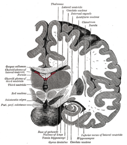 Coronal section of brain immediately in front of pons.
 Coronal section of brain through intermediate mass of third ventricle.
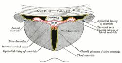 Coronal section of lateral and third ventricles.
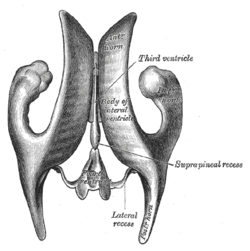 Drawing of a cast of the ventricular cavities, viewed from above.
 Coronal section of brain through anterior commissure.
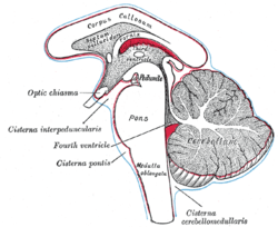 Diagram showing the positions of the three principal subarachnoid cisternæ.
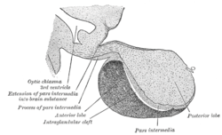 Median sagittal through the hypophysis of an adult monkey. Semidiagrammatic.
 Third ventricle
 Third ventricle
External links
| Wikimedia Commons has media related to Third ventricle. |