Supraspinatous fossa
| Supraspinatous fossa | |
|---|---|
 Left scapula. Dorsal surface. Supraspinatous fossa shown in red. | |
 Left scapula. Dorsal surface. Supraspinatous fossa shown in red. | |
| Details | |
| Identifiers | |
| Latin | fossa supraspinata |
| TA | A02.4.01.007 |
| FMA | 23269 |
The supraspinatous fossa (supraspinatus fossa, supraspinous fossa) of the posterior aspect of the scapula is smaller than the infraspinatous fossa, concave, smooth, and broader at its vertebral than at its humeral end. Its medial two-thirds give origin to the Supraspinatus.
Structure
The fossa can be exposed by the removal of skin and the superficial fascia of the back and the trapezius muscle.
The supraspinatous fossa is bounded by the spine of scapula on the inferior side, acromion process on the lateral side and the superior angle of scapula on the superior side.
Supraspinatus muscle originates from the supraspinatous fossa. Distal attachment of the levator scapulae muscle is also on the medial aspect of the fossa.
Blood supply and innervation
Suprascapular artery and nerve are found within the fossa. The posterior branch of the suprascapular artery supplies the supraspinatous muscle. Dorsal scapular artery also gives off a collateral branch and anastomoses with the suprascapular artery.[1] Suprascapular nerve from the brachial plexus passes through the suprascapular notch as it approaches the fossa to supply the supraspinatus muscle. Suprascapular artery and nerve descend together but are separated by the superior transverse scapular ligament at the suprascapular notch.
Clinical significance
Rotator cuff tear
Hollowing in the supraspinatous and the infraspinatous area is frequently seen as chronic rotator cuff tear resulting in wasting.[2] The wasting may be caused by the supraglenoid cyst compressing the suprascapular nerve and causes a loss of innervation to supraspinatus and infraspinatus muscles. Such wasting or hollowing can be differentially diagnosed as nerve compression or tendon rupture.
Additional images
 The human scapula
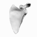 Supraspinatous fossa shown in red.
 Supraspinatous fossa shown in red.
See also
References
- ↑ Clinically Oriented Anatomy (7th ed.). LWW. 2013-02-13. ISBN 9781451119459.
- ↑ Loudon, Janice Kaye; Manske, Robert C.; Reiman, Michael P. (2013-01-01). Clinical Mechanics and Kinesiology. Human Kinetics. ISBN 9780736086431.
This article incorporates text in the public domain from the 20th edition of Gray's Anatomy (1918)
External links
| Wikimedia Commons has media related to Supraspinatous fossa. |
- Anatomy figure: 03:01-02 at Human Anatomy Online, SUNY Downstate Medical Center