Stria vascularis of cochlear duct
| Stria vascularis of cochlear duct | |
|---|---|
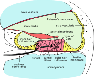 Cross section of the cochlea. | |
| Details | |
| Identifiers | |
| Latin | stria vascularis ductus cochlearis |
| MeSH | A09.246.631.246.292.876 |
| TA | A15.3.03.096 |
| FMA | 77832 |
The upper portion of the spiral ligament (which forms the outer wall of the cochlear duct) contains numerous capillary loops and small blood vessels, and is termed the stria vascularis. It produces endolymph for the scala media, one of the three fluid-filled compartments of the cochlea. The stria is a somewhat stratified epithelium containing primarily three cell types (marginal, intermediate, and basal cells) and intraepithelial capillaries. The marginal cells are involved primarily in K+ transport and line the endolymphatic space of the scala media. The intermediate pigment-containing cells are scattered among capillaries. The basal cells separate stria vascularis from the underlying spiral ligament.[1] The stria vascularis also contains pericyte, melanocyte, and endothelial cells.[2] It is the only epithelial tissue that is not avascular (i.e. completely lacking blood and lymphatic vessels).
References
This article incorporates text in the public domain from the 20th edition of Gray's Anatomy (1918)
- ↑ Ross, Michael H. Histology : a text and atlas / Michael H. Ross, Wojech Pawlina., -6th ed. p 940.
- ↑ Cummings (2001). "Chapter 140: Cochlear Anatomy and Central Auditory Pathways". Stria vascularis (PDF). Textbook of Otolaryngology. p. 3. Retrieved 14 February 2011.