Sebaceous cyst
| Sebaceous cyst | |
|---|---|
 | |
| Pronunciation | /sɪˈbeɪʃəs sɪst/ |
| Classification and external resources | |
| Specialty | Dermatology, general surgery |
| ICD-10 |
Epidermoid cyst L72.0, Pilar cyst L72.1 |
| ICD-9-CM | 706.2 |
| DiseasesDB | 29388 |
| MedlinePlus | 000842 |
| MeSH | D004814 |
A sebaceous cyst is a term commonly used to refer to either:[1]
- Epidermoid cysts (also termed epidermal cysts, infundibular cyst), or
- Pilar cysts (also termed trichelemmal cysts, isthmus-catagen cysts).
Both of the above types of cyst contain keratin, not sebum, and neither originates from sebaceous glands. Epidermoid cysts originate in the epidermis and pilar cysts originate from hair follicles. Therefore, technically speaking they are not sebaceous cysts.[2] "True" sebaceous cysts, cysts which originate from sebaceous glands and which contain sebum, are relatively rare and are known as steatocystoma simplex or, if multiple, as steatocystoma multiplex.
It has been suggested by medical professionals that the term sebaceous cyst be avoided since it can be misleading.[3]:31 In practice however, the term is still often used for epidermoid and pilar cysts.
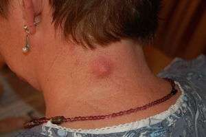
Signs and symptoms

The scalp, ears, back, face, and upper arm, are common sites of sebaceous cysts, though they may occur anywhere on the body except the palms of the hands and soles of the feet. In males a common place for them to develop is the scrotum and chest. They are more common in hairier areas, where in cases of long duration they could result in hair loss on the skin surface immediately above the cyst. They are smooth to the touch, vary in size, and are generally round in shape.
They are generally mobile masses that can consist of:
- Fibrous tissues and fluids,
- A fatty (keratinous) substance that resembles cottage cheese, in which case the cyst may be called "keratin cyst". This material has a characteristic "cheesy" or foot odor smell,
- A somewhat viscous, serosanguineous fluid (containing purulent and bloody material).
The nature of the contents of a sebaceous cyst, and of its surrounding capsule, differs depending on whether the cyst has ever been infected.
With surgery, a cyst can usually be excised in its entirety. Poor surgical technique or previous infection leading to scarring and tethering of the cyst to the surrounding tissue may lead to rupture during excision and removal. A completely removed cyst will not recur, though if the patient has a predisposition to cyst formation, further cysts may develop in the same general area.
Causes
Blocked sebaceous glands, swollen hair follicles,[4] high levels of testosterone and the use of androgenic anabolic steroids[5] will cause such cysts.[6]
A case has been reported of a sebaceous cyst being caused by the human botfly.[7]
Hereditary causes of sebaceous cysts include Gardner's syndrome and basal cell nevus syndrome.
Types
Epidermoid cyst
Pilar cyst
About 90% of pilar cysts occur on the scalp, with the remaining sometimes occurring on the face, trunk and extremities.[8]:1477 Pilar cysts are significantly more common in females, and a tendency to develop these cysts is often inherited in an autosomal dominant pattern.[8]:1477 In most cases, multiple pilar cysts appear at once.[8]:1477
Treatment
Sebaceous cysts generally do not require medical treatment. However, if they continue to grow, they may become unsightly, painful, infected, or all of the above.
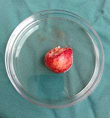
Surgical
Surgical excision of a sebaceous cyst is a simple procedure to completely remove the sac and its contents.[9]
There are three general approaches used: traditional wide excision, minimal excision, and punch biopsy excision.[10]
The typical outpatient surgical procedure for cyst removal is to numb the area around the cyst with a local anaesthetic, then to use a scalpel to open the lesion with either a single cut down the center of the swelling, or an oval cut on both sides of the centerpoint. If the cyst is small, it may be lanced instead. The person performing the surgery will squeeze out the keratin surrounding the cyst, then use blunt-headed scissors or another instrument to hold the incision wide open while using fingers or forceps to try to remove the cyst intact. If the cyst can be removed in one piece, the "cure rate" is 100%. If, however, it is fragmented and cannot be entirely recovered, the operator may use curettage (scraping) to remove the remaining exposed fragments, then burn them with an electro-cauterization tool, in an effort to destroy them in place. In such cases the cyst may recur. In either case, the incision is then disinfected and, if necessary, the skin is stitched back together over it. A scar will most likely result. In some cases where "cure rate" is not 100% the resulting hole is filled with an antiseptic ribbon after washing it with an iodine based solution. This is then covered with a field dressing. The ribbon and the dressing are to be changed once or twice daily for 7–10 days after which the incision is sewn up or allowed to close by secondary intention, i.e. by forming granulation tissue and healing "from the bottom up."
An infected cyst may require oral antibiotics or other treatment before or after excision. If pus has already formed then incision and drainage should be done along with avulsion of cyst wall with proper antibiotics coverage.
An approach involving incision, rather than excision, has also been proposed.[11]
References
- ↑ "Epidermoid and pilar cysts (previously known as sebaceous cysts)". British Association of Dermatologists. Retrieved April 2, 2014.
- ↑ "Epidermoid and Pilar Cysts (Sebaceous Cysts) - Patient UK". Retrieved 2013-03-04.
- ↑ Neville BW, Damm DD, Allen CA, Bouquot JE (2002). Oral & maxillofacial pathology (2nd ed.). Philadelphia: W.B. Saunders. ISBN 0721690033.
- ↑ MedlinePlus Encyclopedia Sebaceous cyst
- ↑ Scott, MJ; Scott, AM (1992). "Effects of anabolic-androgenic steroids on the pilosebaceous unit". Cutis. 50 (2): 113–116. PMID 1387354. Retrieved 30 November 2014.
- ↑ Zuber TJ (2002). "Minimal excision technique for epidermoid (sebaceous) cysts". Am Fam Physician. 65 (7): 1409–12, 1417–8, 1420. PMID 11996426.
- ↑ Harbin LJ, Khan M, Thompson EM, Goldin RD (2002). "A sebaceous cyst with a difference: Dermatobia hominis". J. Clin. Pathol. 55 (10): 798–9. doi:10.1136/jcp.55.10.798. PMC 1769786
 . PMID 12354816.
 - 1 2 3 Barnes L, ed. (2008). Surgical pathology of the head and neck (3rd ed.). New York: Informa Healthcare. ISBN 978-0849390234.
- ↑ Klin B, Ashkenazi H (1990). "Sebaceous cyst excision with minimal surgery". American Family Physician. 41 (6): 1746–8. PMID 2349906.
- ↑ Moore RB, Fagan EB, Hulkower S, Skolnik DC, O'Sullivan G (2007). "Clinical inquiries. What's the best treatment for sebaceous cysts?". The Journal of family practice. 56 (4): 315–6. PMID 17403333.
- ↑ Nakamura M (2001). "Treating a sebaceous cyst: an incisional technique". Aesthetic plastic surgery. 25 (1): 52–6. doi:10.1007/s002660010095. PMID 11322399.
External links
| Wikimedia Commons has media related to Sebaceous cysts. |
- Overview at University of Maryland Medical Center
- Epidermal Inclusion Cyst at eMedicine