Refractive error
| Refraction error | |
|---|---|
|
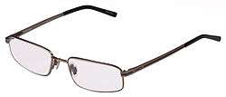
Glasses are a common treatment for refractive errors | |
| Classification and external resources | |
| Specialty | ophthalmology |
| ICD-10 | H52.0-H52.4 |
| ICD-9-CM | 367.0-367.2-367.9 |
| DiseasesDB | 29645 |
| MeSH | D012030 |
Refractive error, also known as refraction error, is a problem with focusing of light on the retina due to the shape of the eye.[1] The most common types of refractive error are near-sightedness, far-sightedness, astigmatism, and presbyopia. Near-sightedness results in far objects being blurry, far-sightedness result in close objects being blurry, astigmatism causes objects to appear stretched out or blurry, and presbyopia results in a poor ability to focus on close objects. Other symptoms may include double vision, headaches, and eye strain.[1]
Near-sightedness is due to the length of the eyeball being too long, far-sightedness the eyeball too short, astigmatism the cornea being the wrong shape, and presbyopia aging of the lens of the eye such that it cannot change shape sufficiently. Some refractive errors are inherited from a person's parents. Diagnosis is by eye examination.[1]
Refractive errors are corrected with eyeglasses, contact lenses, or surgery. Eyeglasses are the easiest and safest method of correction. Contact lenses can provide a wider field of vision; however are associated with a risk of infection. Refractive surgery permanently changes the shape of the cornea.[1]
The number of people globally with refractive errors has been estimated at one to two billion. Rates vary between regions of the world with about 25% of Europeans and 80% of Asians affected.[2] Near-sightedness is the most common disorder.[3] Rates among adults are between 15-49% while rates among children are between 1.2-42%.[4] Far-sightedness more commonly affects young child and the elderly.[5][6] Presbyopia affects most people over the age of 35.[1] The number of people with refractive errors that have not been corrected was estimated at 660 million (10 per 100 people) in 2013.[7] Of these 9.5 million were blind due to the refractive error.[7] It is one of the most common causes of vision loss along with cataracts, macular degeneration, and vitamin A deficiency.[8]
Classification
An eye that has no refractive error when viewing distant objects is said to have emmetropia or be emmetropic meaning the eye is in a state in which it can focus parallel rays of light (light from distant objects) on the retina, without using any accommodation. A distant object in this case is defined as an object 8 meters or further away from the eye.
An eye that has refractive error when viewing distant objects is said to have ametropia or be ametropic. This eye cannot focus parallel rays of light (light from distant objects) on the retina, or needs accommodation to do so.
The word "ametropia" can be used interchangeably with "refractive error". Types of ametropia include myopia, hyperopia and astigmatism. They are frequently categorized as spherical errors and cylindrical errors:
- Spherical errors occur when the optical power of the eye is either too large or too small to focus light on the retina. People with refractive error frequently have blurry vision.
- Nearsightedness: When the optics are too powerful for the length of the eyeball one has myopia or nearsightedness. This can arise from a cornea or crystalline lens with too much curvature (refractive myopia) or an eyeball that is too long (axial myopia). Myopia can be corrected with a concave lens which causes the divergence of light rays before they reach the cornea.
- Farsightedness: When the optics are too weak for the length of the eyeball, one has hyperopia or farsightedness. This can arise from a cornea or crystalline lens with not enough curvature (refractive hyperopia) or an eyeball that is too short (axial hyperopia). This can be corrected with convex lenses which cause light rays to converge prior to hitting the cornea.
- Presbyopia: When the flexibility of the lens declines, typically due to age. The individual would experience difficulty in near vision, often relieved by reading glasses, bifocal, or progressive lenses.
- Cylindrical errors cause astigmatism, when the optical power of the eye is too powerful or too weak across one meridian, such as if the corneal curvature tends towards a cylindrical shape. The angle between that meridian and the horizontal is known as the axis of the cylinder.
- Astigmatism: A person with astigmatic refractive error sees lines of a particular orientation less more clearly than lines at right angles to them. This defect can be corrected by refracting light more in one meridian than the other. Cylindrical lenses serve this purpose.
Risk factors
Genetics
The Online Mendelian Inheritance in Man (OMIM) database has listed 261 genetic disorders in which myopia is one of the symptoms.[9] Myopia may be present in heritable connective tissue disorders such as: Knobloch syndrome (OMIM 267750); Marfan syndrome (OMIM 154700); and Stickler syndrome (type 1, OMIM 108300; type 2, OMIM 604841).[10] Myopia is present in heritable connective tissue disorders such as: Knobloch syndrome (OMIM 267750); Marfan syndrome (OMIM 154700); and Stickler syndrome (type 1, OMIM 108300; type 2, OMIM 604841).[10] Myopia has also been reported in X-linked disorders caused by mutations in loci involved in retinal photoreceptor function (NYX, RP2, MYP1) such as: autosomal recessive congenital stationary night blindness (CSNB; OMIM 310500); retinitis pigmentosa 2 (RP2; OMIM 312600); Bornholm eye disease (OMIM 310460).[11] Many genes that have been associated with refractive error are clustered into common biological networks involved in connective tissue growth and extracellular matrix organization.[10] Although a large number of chromosomal localisations have been associated with myopia (MYP1-MYP17), few specific genes have been identified.[9]
Environmental
In studies of the genetic predisposition of refractive error, there is a correlation between environmental factors and the risk of developing myopia.[12] Myopia has been observed in individuals with visually intensive occupations.[11] Reading has also been found to be a predictor of myopia in children. It has been reported that children with myopia spent significantly more time reading than non-myopic children who spent more time playing outdoors.[11] Socioeconomic status and higher levels of education have also been reported to be a risk factor for myopia.
Diagnosis
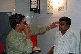
Blurry vision may result from any number of conditions not necessarily related to refractive errors. The diagnosis of a refractive error is usually confirmed by an eye care professional during an eye examination using a large number of lenses of different optical powers, and often a retinoscope (a procedure entitled retinoscopy) to measure objectively in which the patient views a distant spot while the clinician changes the lenses held before the patient's eye and watches the pattern of reflection of a small light shone on the eye. Following that "objective refraction" the clinician typically shows the patient lenses of progressively higher or weaker powers in a process known as subjective refraction. Cycloplegic agents are frequently used to more accurately determine the amount of refractive error, particularly in children[13]
An automated refractor is an instrument that is sometimes used in place of retinoscopy to objectively estimate a person's refractive error.[14] Shack–Hartmann wavefront sensor and its inverse[15] can also be used to characterize eye aberrations in a higher level of resolution and accuracy.
Vision defects caused by refractive error can be distinguished from other problems using a pinhole occluder, which will improve vision only in the case of refractive error.
Management
How refractive errors are treated or managed depends upon the amount and severity of the condition. Those who possess mild amounts of refractive error may elect to leave the condition uncorrected, particularly if the patient is asymptomatic. For those who are symptomatic, glasses, contact lenses, refractive surgery, or a combination of the three are typically used.
In the case of myopia, however, some believe that such treatments may also have the long-term effect of exacerbating that refractive error — i.e., making the patient even more nearsighted. This would be due to the very same prescription that is tailored for use at a 12-to-20-foot distance also commonly being used for close-up work as well, thus artificially amplifying the focusing stress that would normally be presented to the accommodation mechanisms of the eye at that distance.
However, this exacerbating effect is not generally believed to exist in the general case, although in cases where the myopia is due to accommodative spasm, removing the corrective lenses for a time may lead to improvement.
Epidemiology

The number of people globally with refractive errors that have not been corrected was estimated at 660 million (10 per 100 people) in 2013.[7]
The number of people globally with refractive errors has been estimated from 800 million to 2.3 billion.
References
- 1 2 3 4 5 "Facts About Refractive Errors". NEI. October 2010. Retrieved 29 July 2016.
- ↑ Denniston, Alastair; Murray, Philip (2014). Oxford Handbook of Ophthalmology (3 ed.). OUP Oxford. p. 826. ISBN 9780191057021.
- ↑ Foster, PJ; Jiang, Y (February 2014). "Epidemiology of myopia.". Eye (London, England). 28 (2): 202–8. PMID 24406412.
- ↑ Pan, CW; Ramamurthy, D; Saw, SM (January 2012). "Worldwide prevalence and risk factors for myopia.". Ophthalmic & physiological optics : the journal of the British College of Ophthalmic Opticians (Optometrists). 32 (1): 3–16. PMID 22150586.
- ↑ Castagno, VD; Fassa, AG; Carret, ML; Vilela, MA; Meucci, RD (23 December 2014). "Hyperopia: a meta-analysis of prevalence and a review of associated factors among school-aged children.". BMC ophthalmology. 14: 163. PMID 25539893.
- ↑ Grosvenor, Theodore (2007). Primary care optometry (5 ed.). St. Louis (Miss.): Butterworth Heinemann, Elsevier. p. 70. ISBN 9780750675758.
- 1 2 3 Global Burden of Disease Study 2013, Collaborators (22 August 2015). "Global, regional, and national incidence, prevalence, and years lived with disability for 301 acute and chronic diseases and injuries in 188 countries, 1990-2013: a systematic analysis for the Global Burden of Disease Study 2013.". Lancet (London, England). 386 (9995): 743–800. PMID 26063472.
- ↑ Pan, CW; Dirani, M; Cheng, CY; Wong, TY; Saw, SM (March 2015). "The age-specific prevalence of myopia in Asia: a meta-analysis.". Optometry and vision science : official publication of the American Academy of Optometry. 92 (3): 258–66. PMID 25611765.
- 1 2 Morgan, Ian; Kyoko Ohno-Matsui (May 2012). "Myopia". The Lancet. 379 (9827): 1739–1748. doi:10.1016/S0140-6736(12)60272-4.
- 1 2 3 Wojciechowski, Robert (April 2011). "Nature and Nurture: the complex genetics of myopia and refractive error". Clin Genet. 79 (4): 301–320. doi:10.1111/j.1399-0004.2010.01592.x.
- 1 2 3 Wojcienchowski, Robert (April 2011). "Nature and Nurture: the complex genetics of myopia and refractive error". National Institutes of Health. 79 (4): 301–320. doi:10.1111/j.1399-0004.2010.01592.x.
- ↑ Barnes, Katherine (February 2013). "Genome-wide meta-analyses of multiancestry cohorts identify multiple new susceptibility loci for refractive error and myopia". Nature Genetics. 45 (3): 314–8. doi:10.1038/ng.2554. PMC 3740568
 . PMID 23396134.
 - ↑ Roque, B. Refractive errors in children. November 2, 2005.
- ↑ "Frequently Asked Questions: How do you measure refractive errors?". The New York Eye And Ear Infirmary. Retrieved 2006-09-13.
- ↑ "NETRA: Inverse Shack-Hartmann Wavefront Sensor using High Resolution Mobile Phone Display". Vitor F. Pamplona, Ankit Mohan, Manuel M. Oliveira, Ramesh Raskar. Retrieved 2011-12-13.
- ↑ "WHO Disease and injury country estimates". World Health Organization. 2009. Retrieved Nov 11, 2009.