cGMP-dependent protein kinase
| protein kinase, cGMP-dependent, type I | |
|---|---|
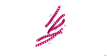 Crystallographic structure of the leucine zipper domain of human cGMP dependent protein kinase I beta.[1] | |
| Identifiers | |
| Symbol | PRKG1 |
| Alt. symbols | PRKGR1B, PRKG1B |
| Entrez | 5592 |
| HUGO | 9414 |
| OMIM | 176894 |
| RefSeq | NM_006258 |
| UniProt | Q13976 |
| Other data | |
| Locus | Chr. 10 q11.2 |
| protein kinase, cGMP-dependent, type II | |
|---|---|
| Identifiers | |
| Symbol | PRKG2 |
| Entrez | 5593 |
| HUGO | 9416 |
| OMIM | 601591 |
| RefSeq | NM_006259 |
| UniProt | Q13237 |
| Other data | |
| Locus | Chr. 4 q13.1-21.1 |
cGMP-dependent protein kinase or Protein Kinase G (PKG) is a serine/threonine-specific protein kinase that is activated by cGMP. It phosphorylates a number of biologically important targets and is implicated in the regulation of smooth muscle relaxation, platelet function, sperm metabolism, cell division, and nucleic acid synthesis.
Genes and proteins
PKG are serine/threonine kinases that are present in a variety of eukaryotes ranging from the unicellular organism Paramecium to humans. Two PKG genes, coding for PKG type I (PKG-I) and type II (PKG-II), have been identified in mammals. The N-terminus of PKG-I is encoded by two alternatively spliced exons that specify for the PKG-Iα and PKG-Iβ isoforms. PKG-Iβ is activated at ~10-fold higher cGMP concentrations than PKG-Iα. The PKG-I and PKG-II are homodimers of two identical subunits (~75 kDa and ~85 kDa, respectively) and share common structural features.
Each subunit is composed of three functional domains:
- (1) an N-terminal domain that mediates homodimerization, suppression of the kinase activity in the absence of cGMP, and interactions with other proteins including protein substrates
- (2) a regulatory domain that contains two non-identical cGMP-binding sites
- (3) a kinase domain that catalyzes the phosphate transfer from ATP to the hydroxyl group of a serine/threonine side chain of the target protein
Binding of cGMP to the regulatory domain induces a conformational change which stops the inhibition of the catalytic core by the N-terminus and allows the phosphorylation of substrate proteins. Whereas PKG-I is predominantly localized in the cytoplasm, PKG-II is anchored to the plasma membrane by N-terminal myristoylation.
Tissue distribution
In general, PKG-I and PKG-II are expressed in different cell types.
- PKG-I has been detected at high concentrations (above 0.1 µmol/L) in all types of smooth muscle cells (SMCs) including vascular SMCs and in platelets. Lower levels are present in vascular endothelium and cardiomyocytes. The enzyme is also expressed in fibroblasts, certain types of renal cells and leukocytes, and in specific regions of the nervous system, for example in the hippocampus, in cerebellar Purkinje cells, and in dorsal root ganglia. Neurons express either the PKG-Iα or the PKG-Iβ isoform, platelets predominantly Iβ, and both isoforms are present in smooth muscle.
- PKG-II has been detected in renal cells, zona glomerulosa cells of the adrenal cortex, club cells in distal airways, intestinal mucosa, pancreatic ducts, parotid and submandibular glands, chondrocytes, and several brain nuclei, but not in cardiac and vascular myocytes.
Specifically, in smooth muscle tissue, PKG promotes the opening of calcium-activated potassium channels,[2] leading to cell hyperpolarization and relaxation, and blocks agonist activity of phospholipase C, reducing liberation of stored calcium ions by inositol triphosphate.
Role in cancer
Cancerous colon cells stop producing PKG, which apparently limits beta-catenin thus allowing the VEGF enzyme to solicit angiogenesis.[3]
See also
References
- ↑ PDB: 3NMD; Casteel DE, Smith-Nguyen EV, Sankaran B, Roh SH, Pilz RB, Kim C (October 2010). "A crystal structure of the cyclic GMP-dependent protein kinase I{beta} dimerization/docking domain reveals molecular details of isoform-specific anchoring". J. Biol. Chem. 285 (43): 32684–8. doi:10.1074/jbc.C110.161430. PMC 2963381
 . PMID 20826808.
 - ↑ Kyle BD, Hurst S, Swayze RD, Sheng J, Braun AP (Feb 2013). "Specific phosphorylation sites underlie the stimulation of a large conductance, Ca(2+)-activated K(+) channel by cGMP-dependent protein kinase". The FASEB Journal. 27 (5): 2027–38. doi:10.1096/fj.12-223669. PMID 23407708.
- ↑ Kwon IK, Schoenlein PV, Delk J, Liu K, Thangaraju M, Dulin NO, Ganapathy V, Berger FG, Browning DD (April 2008). "Expression of cyclic guanosine monophosphate-dependent protein kinase in metastatic colon carcinoma cells blocks tumor angiogenesis". Cancer. 112 (7): 1462–70. doi:10.1002/cncr.23334. PMID 18260092.
External links
- EC 2.7.11.12
- Cyclic GMP-Dependent Protein Kinases and the Cardiovascular System
- cGMP-Dependent Protein Kinases at the US National Library of Medicine Medical Subject Headings (MeSH)