Polymyxin

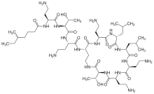
Polymyxins are antibiotics,[1] with a general structure consisting of a cyclic peptide with a long hydrophobic tail. They disrupt the structure of the bacterial cell membrane by interacting with its phospholipids. They are produced by nonribosomal peptide synthetase systems in Gram-positive bacteria such as Paenibacillus polymyxa[2] and are selectively toxic for Gram-negative bacteria due to their specificity for the lipopolysaccharide molecule that exists within many Gram-negative outer membranes.
Polymyxins B and E (also known as colistin) are used in the treatment of Gram-negative bacterial infections. The global problem of advancing antimicrobial resistance has led to a renewed interest in their use recently.[3]
Polymyxin M is also known as "mattacin".[4]
Clinical use
Polymyxin antibiotics are relatively neurotoxic and nephrotoxic,[5] so are usually used only as a last resort if modern antibiotics are ineffective or are contraindicated. Typical uses are for infections caused by strains of multiple drug-resistant Pseudomonas aeruginosa or carbapenemase-producing Enterobacteriaceae.
Polymyxins B are not absorbed from the gastrointestinal tract, so another route of administration must be chosen, e.g., parenteral (often intravenously) or by inhalation (unless perhaps the targets are bacteria in the gastrointestinal tract). They are also used externally as a cream or drops to treat otitis externa (swimmers ear).[6] and as a component of triple antibiotic ointment to treat and prevent skin infections.
Polymyxins have less effect on Gram-positive organisms, and are sometimes combined with other agents (as with trimethoprim/polymyxin) to broaden the effective spectrum.
Mechanism of action
After binding to lipopolysaccharide (LPS) in the outer membrane of Gram-negative bacteria, polymyxins disrupt both the outer and inner membranes. The hydrophobic tail is important in causing membrane damage, suggesting a detergent-like mode of action.[7]
Removal of the hydrophobic tail of polymyxin B yields polymyxin nonapeptide, which still binds to LPS, but no longer kills the bacterial cell. However, it still detectably increases the permeability of the bacterial cell wall to other antibiotics, indicating it still causes some degree of membrane disorganization.[8]
Gram-negative bacteria can develop resistance to polymyxins through various modifications of the LPS structure that inhibit the binding of polymyxins to LPS.[9]
Antibiotic resistance to this drug has been increasing, especially in southern China. Recently the gene mcr-1, which confers the antibiotic resistance, has been isolated from Enterobacteriaceae bacteria plasmids which will increase the likelihood of its spread globally.[10][11]
Use in biomedical research
Polymyxins are used to neutralize or absorb LPS, which contaminates samples intended for use in, e.g., immunological experiments. Minimization of LPS contamination can be important because LPS can evoke strong reactions from immune cells and, therefore, distort experimental results.
By increasing permeability of the bacterial membrane system, polymyxin is also used in clinical work to increase release of secreted toxins, such as Shiga toxin from Escherichia coli.[12]
See also
References
- ↑ Dixon RA, Chopra I (November 1986). "Polymyxin B and polymyxin B nonapeptide alter cytoplasmic membrane permeability in Escherichia coli". J. Antimicrob. Chemother. 18 (5): 557–563. doi:10.1093/jac/18.5.557. PMID 3027012.
- ↑ Polymyxins at the US National Library of Medicine Medical Subject Headings (MeSH)
- ↑ Falagas ME, Grammatikos AP, Michalopoulos A. Potential of old-generation antibiotics to address current need for new antibiotics. Expert Rev Anti Infect Ther. 2008; 6(5):593-600 PMID 18847400
- ↑ Martin NI, Hu H, Moake MM, et al. (April 2003). "Isolation, structural characterization, and properties of mattacin (polymyxin M), a cyclic peptide antibiotic produced by Paenibacillus kobensis M". J. Biol. Chem. 278 (15): 13124–13132. doi:10.1074/jbc.M212364200. PMID 12569104.
- ↑ Falagas ME, Kasiakou SK (February 2006). "Toxicity of polymyxins: a systematic review of the evidence from old and recent studies". Crit Care. 10 (1): R27. doi:10.1186/cc3995. PMC 1550802
 . PMID 16507149.
 - ↑ Staff, Medicine.net. Last reviewed 16 April 2014 Neomycin/polymyxin/hydrocortisone suspension - otic, Cortisporin, Pediotic
- ↑ Velkov, Tony; Roberts, Kade D; Nation, Roger L; Thompson, Philip E; Li, Jian (2013-06-01). "Pharmacology of polymyxins: new insights into an 'old' class of antibiotics". Future microbiology. 8 (6). doi:10.2217/fmb.13.39. ISSN 1746-0913. PMC 3852176
 . PMID 23701329.
 - ↑ Tsubery, H.; Ofek, I.; Cohen, S.; Fridkin, M. (2000-01-01). "Structure activity relationship study of polymyxin B nonapeptide". Advances in Experimental Medicine and Biology. 479: 219–222. doi:10.1007/0-306-46831-X_18. ISSN 0065-2598. PMID 10897422.
- ↑ Tran AX, Lester ME, Stead CM, et al. (August 2005). "Resistance to the antimicrobial peptide polymyxin requires myristoylation of Escherichia coli and Salmonella typhimurium lipid A". J. Biol. Chem. 280 (31): 28186–28194. doi:10.1074/jbc.M505020200. PMID 15951433.
- ↑ "Antibiotic resistance threatens the efficacy of prophylaxis - The Lancet Infectious Diseases". thelancet.com. Retrieved 2015-11-19.
- ↑ Liu YY; et al. (18 November 2015). "Emergence of plasmid-mediated colistin resistance mechanism MCR-1 in animals and human beings in China: a microbiological and molecular biological study". The Lancet. doi:10.1016/S1473-3099(15)00424-7.
- ↑ Yokoyama et al. 2000. FEMS Microbiol. Lett. 192:139-144.
Further reading
- Giuliani A, Pirri G, Nicoletto S (2007). "Antimicrobial peptides: an overview of a promising class of therapeutics". Cent. Eur. J. Biol. 2 (1): 1–33. doi:10.2478/s11535-007-0010-5.
- Pirri G, Giuliani A, Nicoletto S, Pizutto L, Rinaldi A (2009). "Lipopeptides as anti-infectives: a practical perspective". Cent. Eur. J. Biol. 4 (3): 258–273. doi:10.2478/s11535-009-0031-3.
