Plastocyanin
Plastocyanin is a copper-containing protein involved in electron-transfer. The protein is a monomer, with a molecular weight around 10,500 Daltons, and 99 amino acids in most vascular plants. It is a member of the plastocyanin family of copper-binding proteins.
Function
In photosynthesis, plastocyanin functions as an electron transfer agent between cytochrome f of the cytochrome b6f complex from photosystem II and P700+ from photosystem I. Cytochrome b6f complex and P700+ are both membrane-bound proteins with exposed residues on the lumen-side of the thylakoid membrane of chloroplasts. Cytochrome f acts as an electron donor while P700+ accepts electrons from reduced plastocyanin.
Structure
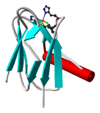
Plastocyanin was the first of the blue copper proteins to be characterised by X-ray crystallography. The tertiary structure is a beta-barrel — common in proteins which bind to other proteins.[1]
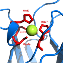
Although the molecular surface of plastocyanins differs for plants, algae, and cyanobacteria, the structure of the copper binding site is generally conserved. The geometry of the copper binding site is described as a ‘distorted trigonal pyramidal’. The trigonal plane of the pyramidal base is composed of two nitrogen atoms (N1 and N2) from separate histidine residues and a sulfur atom (S1) from a cysteine residue. A second sulfur atom (S2) from an axial methionine residue forms the apex. The ‘distortion’ occurs in the bond lengths between the copper atom and sulfur ligands. The Cu-S1 contact is much shorter (207 picometers) than Cu-S2 (282 pm). The elongated Cu-S2 bonding destabilises the CuII form and increases the redox potential of the protein. The blue colour (597 nm peak absorption) is due to the Cu-S1 bond where Spπ to Cudx2-y2 charge transfer occurs.[2]
In the reduced form of plastocyanin, His-87 will become protonated with a pKa of 4.4. Protonation prevents it acting as a ligand and the copper site geometry becomes trigonal planar.
While the molecular surface of the protein near the copper binding site varies slightly, all plastocyanins have a hydrophobic surface surrounding the exposed histidine of the copper binding site. In plant plastocyanins, acidic residues are located on either side of the highly conserved tyrosine-83. Algal plastocyanins, and those from vascular plants in the family Apiaceae, contain similar acidic residues but are shaped differently from those of plant plastocyanins—they lack residues 57 and 58. In cyanobacteria, the distribution of charged residues on the surface is different from eukaryotic plastocyanins and variations among different bacterial species is large. Many cyanobacterial plastocyanins have 107 amino acids. Although the acidic patches are not conserved in bacteria the hydrophobic patch is always present. These hydrophobic and acidic patches are believed to be the recognition/binding sites for the other proteins involved in electron transfer.
Reactions
Plastocyanin (Cu2+Pc) is reduced (an electron is added) by cytochrome f according to the following reaction:
- Cu2+Pc + e− → Cu+Pc
After dissociation, Cu+Pc diffuses through the lumen space until recognition/binding occurs with P700+, at which point P700+ oxidizes Cu+Pc according to the following reaction:
- Cu+Pc → Cu2+Pc + e−
The redox potential is about 370 mV[3] and the isoelectric pH is about 4.[4]
References
Notes
- ↑ Freeman, H. C.; Guss, J. M. (2001). "Plastocyanin". In Bode, Wolfram; Messerschmidt, Albrecht; Cygler, Mirek. Handbook of metalloproteins. 2. Chichester: John Wiley & Sons. pp. 1153–69. ISBN 0-471-62743-7.
- ↑ Gewirth AA, Solomon EI (June 1988). "Electronic structure of plastocyanin: excited state spectral features". J Am Chem Soc. 110: 3811–9. doi:10.1021/ja00220a015.
- ↑ Anderson GP, Sanderson DG, Lee CH, Durell S, Anderson LB, Gross EL (December 1987). "The effect of ethylenediamine chemical modification of plastocyanin on the rate of cytochrome f oxidation and P-700+ reduction". Biochim. Biophys. Acta. 894 (3): 386–98. doi:10.1016/0005-2728(87)90117-4. PMID 3689779.
- ↑ Ratajczak R, Mitchell R, Haehnel W (1988). "Properties of the oxidizing site of Photosystem I". Biochim. Biophys. Acta. 933: 306–318. doi:10.1016/0005-2728(88)90038-2.
Sources
- Berg, Jeremy Mark; Lippard, Stephen J. (1994). "Blue Copper Proteins". Principles of bioinorganic chemistry. Sausalito, Calif: University Science Books. pp. 237–242. ISBN 0-935702-72-5.
- Sato K, Kohzuma T, Dennison C (February 2003). "Active-site structure and electron-transfer reactivity of plastocyanins". J. Am. Chem. Soc. 125 (8): 2101–12. doi:10.1021/ja021005u. PMID 12590538.