Place cell
A place cell is a type of pyramidal neuron within the hippocampus that becomes active when an animal enters a particular place in its environment; this place is known as the place field. A given place cell will have only one, or a few, place fields in a typical small laboratory environment, but more in a larger region.[1] There is no apparent topography to the pattern of place fields, unlike other brain areas such as visual cortex—neighboring place cells are as likely to have nearby fields as distant ones.[2] In a different environment, typically about half the place cells will still have place fields, but these will be in new places unrelated to their former locations.[3]
Place cells are thought, collectively, to act as a cognitive representation of a specific location in space, known as a cognitive map.[4] Place cells work with other types of neurons in the hippocampus and surrounding regions to perform this kind of spatial processing,[5] but the ways in which they function within the hippocampus are still being researched.[6]
Studies with rats have shown that place cells tend to fire quickly when a rat enters a new, open environment, but outside of a firing field, place cells tend to be relatively inactive.[7] Together place cells are thought to form a "cognitive map" in which they have localized firing patterns called place fields.[8] Place cell firing patterns are often determined by external sensory information and the local environment. Place cells have proven to have the ability to suddenly change their firing pattern from one pattern to another, a phenomenon known as "re-mapping" and though place cells do change according to the external environment, they are stabilized by attractor dynamics which "enable the system to resist small changes in sensory input but respond collectively and coherently to large ones."[8]
Although place cells are part of a non-sensory cortical system, their firing behavior is strongly correlated to sensory input. Place cells fire when an animal is located in parts of the environment known as place fields.[9] These circuits may have important implications for memory, as they provide the spatial context for memories and past experiences.[9] Like many other parts of the brain, place cell circuits are dynamic. They are constantly adjusting and remapping to suit the current location and experience of the brain. Place cells do not work alone to create visuospatial representation; they are a part of a complex circuit that informs place awareness and place memory.[9]
The 2014 Nobel Prize in Physiology or Medicine was awarded to John O'Keefe for the discovery of place cells, and to Edvard and May-Britt Moser for the discovery of grid cells.[10][11]
Background
These cells were first discovered in the brain, and specifically in the hippocampus, by O'Keefe and Dostrovsky (1971).[12] Though the hippocampus plays a role in learning and memory, the existence of place cells within the hippocampus demonstrates the role it plays with spatial adaptation and awareness. There have been recorded increases in firing patterns of rats in open environments and recorded spatial learning and awareness impairments after damage to the hippocampus and the place cells within.[12] Studies with rats have shown that place cells are very responsive to spatial surroundings. For example a study by John O'Keefe and Lynn Nadel found that place cells would fire more rapidly when rats ran past places in the environment, when a new item was added to the environment, or when an item that is usually there is not present.[13]
After O'Keefe and Dostrovsky first found the existence of place cells within the hippocampus in 1971, they conducted a study five years later with rats that demonstrated these place cells would fire whenever the rat was within a certain place in the environment.[14] This was one of the first indicators that place cells were related to spatial orientation. They also discovered that place cells fired in different areas of the hippocampus depending on where the rat went, and this whole firing network made up the rat's environment (O'Keefe 1976, Wilson & McNaughton 1993). As environments changed, the same place cells would fire, but the relationship and dynamic between firing fields would change (O'Keefe & Conway 1978). Therefore place cells are thought to give humans and animals a guide to the environment it is navigating and its position in that environment. Place cells are generally observed through recorded action potentials. As humans or animals navigate large environments and then arrive at a particular location, there is a notable increase in the place cell firing rate once that specific location has been reached (Eichenbaum, Dudchencko Wood, Shaprio and Tanila, 1999). For more information on studies with rats, "Place Cells and Aging".
There has been much debate as to whether hippocampal place cells function based upon landmarks in the environment or on environmental boundaries or an interaction between the two.[15] There has also been much study as to whether hippocampal pyramidal cells (mostly in rats) signal non-spatial information as well as spatial information. According to the cognitive map theory, the hippocampus's primary role in the rat is to store spatial information through place cells and the rat hippocampus was biologically designed to provide the rat with spatial information.[16]
However, there have been investigations as to whether the hippocampus may store other non-spatial information as well.[16] These other explanations in favor of non-spatial components of the hippocampus argue that the hippocampus has "flexible" functions in that it can apply memory in circumstances different from those under which these relationships were learned. There are also views that claim that the hippocampus has functions altogether removed from time and space.[16] However, other explanations of data that prematurely support the existence of non-spatial functions in the hippocampus must be considered. Evidence against this flexibility theory comes in the form of using the delayed non-match-to sample task. This task uses flexibility in that the rat is first presented with a visual representation such as a block. After a delay, when presented with the block and a novel object, the rat must choose the novel object in order to obtain a reward. Its completion of this task requires flexibility. However, during this task, hippocampal activity does not sufficiently increase and lesioning (induced trauma) in the hippocampus does not change the rat's performance on this task.[16]
Place cells fire in different, often widespread, hippocampal locations at the same time, which some interpret as their having different functions in different locations. A rat's representation of its environment is constructed by the firing of groups of place cells that are widely distributed in the hippocampus, however, this does not necessarily mean that each location serves a different purpose. When recording the firing fields of certain hippocampal cells in an open field environment, firing fields prove to be similar even when the rat travels in different directions, exhibiting omnidirectionality. However, when limitations are placed in the aforementioned environment, fields prove to be directional and fire in one direction but not in another.
The same directionality occurs when rats participate in the radial arm maze. The radial arm maze consists of a central circle from which several arm-like projections radiate. These projections either contain food or do not. Some consider the firing or lack of firing of place cells depending on the arm to be a function of goal-oriented behavior. However, when moving from one arm to another when they both contain food, place cells only fire in one direction, meaning that one cannot attribute firing purely to a goal-approach. A directionality component must be added: for example, a North goal as opposed to a South goal.[16]
When visual cues in an environment such as visibility of a line where the wall meets the floor, height of the wall, and width of the wall are available to the rat to discern distance and location of the wall, the rat internalizes this external information to register its surroundings. However, when these visual cues are unavailable, the rat registers wall location by colliding with the wall and then place cell firing rate after the collision provides information to the rat about its distance from the wall based on the direction and speed of its movements after the collision. In this situation, the firing of place cells is due to motor inputs.[16]
There are both simple place cells with purely locational correlates and also complex place cells that increase their firing rate when the rat encounters a particular object or experience. Others fire when a rat's expectations in a particular location are not met or when they encounter novelty along their path: the cells that fire in these situations are known as misplace cells.
The place cells that appear to operate based solely on non-spatial memory seem to have spatial components. Many lesioning experiments attempting to inflict non-spatial memory deficits in the hippocampus have been unsuccessful. In some cases, lesioning has been successful in inflicting non-spatial memory deficits, however, other structures besides the hippocampus were affected by lesioning. Therefore, the rat's non-spatial memory deficits could have been unrelated to place cells.[16] Thus, based on information from studies thus far, the cognitive map theory seems to be most supported and non-spatial theories may fail to take spatial components into account.[16]
Function
Place fields
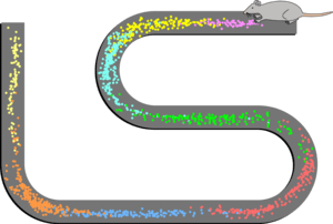
Place cells fire in a specific region known as a place field. Place fields are roughly analogous to the receptive fields of sensory neurons, in that the firing region corresponds to a region of sensory information in the environment. A good depiction of place fields can be seen here[17] This animation shows place fields firing in succession as a rat moves along a linear track. Place fields are thus considered to be allocentric rather than egocentric, meaning that they are defined with respect to the outside world rather than the body. By orienting based on the environment rather than the individual, place fields can work effectively as neural maps of the environment.[18]
Sensory input
Place cells were initially believed to fire in direct relation to simple sensory inputs, but recent studies suggest that this may not be the case.[18] Place fields are usually unaffected by large sensory changes, like removing a landmark from an environment, but respond to subtle changes, like a change in color or shape of an object.[19] This suggests that place cells respond to complex stimuli rather than simple individual sensory cues. According to a model known as the functional differentiation model, sensory information is processed in various cortical structures upstream of the hippocampus before actually reaching the structure, so that the information received by place cells is a compilation of different stimuli.[18] Mechanisms required for memory consolidation affect place cell firing less than direct sensory input does, suggesting that it is primarily recent inputs that are retrieved during place coding.[20]
Sensory information received by place cells can be categorized as either metric or contextual information, where metric information corresponds to where place cells should fire and contextual input corresponds to whether or not a place field should fire in a certain environment.[21] Metric sensory information is any kind of spatial input that might indicate a distance between two points. For example, the edges of an environment might signal the size of the overall place field or the distance between two points within a place field. Metric signals can be either linear or directional. Directional inputs provide information about the orientation of a place field, whereas linear inputs essentially form a representational grid. Contextual cues allow established place fields to adapt to minor changes in the environment, such as a change in object color or shape. Metric and contextual inputs are processed together in the entorhinal cortex before reaching the hippocampal place cells. Visuospatial and olfactory inputs are examples of sensory inputs that are utilized by place cells. These types of sensory cues can include both metric and contextual information.[21]
Visuospatial cues
Spatial cues such as geometric boundaries or orienting landmarks are important examples of metric input. Place cells mainly rely on set distal cues rather than cues in the immediate proximal environment.[21] Movement can also be an important spatial cue. The ability of place cells to incorporate new movement information is called path integration, and it is important for keeping track of self-location during movement.[22] Path integration is largely aided by grid cells, which are a type of neuron in the entorhinal cortex that relay information to place cells in the hippocampus. Grid cells establish a grid representation of a location, so that during movement place cells can fire according to their new location while orienting according to the reference grid of their external environment.[21] Visual sensory inputs can also supply important contextual information. A change in color of a specific object can affect whether or not a place cell fires in a particular field.[21] Thus, visuospatial sensory information is critical to the formation and recollection of place field.
Olfactory cues
Although place cells primarily rely on visuospatial input, some studies suggest that olfactory input may also play a role in generating and recalling place fields.[23][24] Relatively little is known about the interaction between place cells and non-visual sensory cues, but preliminary studies have shown that non-visual sensory input may have supplementary role in place field formation. A study by Save et al. found that olfactory information can be used to compensate for a loss of visual information. In this study, place fields in subjects exposed to an environment with no light and no olfactory signals were unstable; the position of the place field shifted abruptly and some of the constituent place cells stopped firing entirely. However, place cells in subjects exposed to a dark environment with olfactory signals remained stable despite a lack of visual cues.[23] An additional study by Zhang et al. examined how the hippocampus uses olfactory signals to create and recall place fields. Similar to the Save et al. study, this study exposed subjects to an environment with a series of odors but no visual or auditory information. Place fields remained stable and even adapted to the rotation of the pattern of olfactory signals. Furthermore, the place fields would remap entirely when the odors were moved randomly.[24] This suggests that place cells not only utilize olfactory information to generate place fields, but also use olfactory information to orient place fields during movement.
Hippocampal memory
The hippocampus plays an essential role in episodic memory.[25] One important aspect of episodic memory is the spatial context in which the event occurred.[26] Hippocampal place cells have been shown to exhibit stable firing patterns even when cues from a location are removed. Additionally, specific place fields begin firing when exposed to signals or a subset of signals from a previous location.[27] This suggests that place cells provide the spatial context for a memory by recalling the neural representation of the environment in which the memory occurred. In other words, place cells prime a memory by differentiating the context for the event.[26] By establishing spatial context, place cells can be used to complete memory patterns.[25] Furthermore, place cells can maintain a spatial representation of one location while recalling the neural map of a separate location, effectively differentiating between present experience and past memory.[26] Place cells are therefore considered to demonstrate both pattern completion and pattern separation qualities.[25]
Pattern completion
Pattern completion is the ability to recall an entire memory from a partial or degraded sensory cue.[25] Place cells are able to maintain a stable firing field even after significant signals are removed from a location, suggesting that they can recall a pattern from only some of the original input.[19] Furthermore, pattern completion can be symmetric in that an entire memory can be retrieved from any part of it. For example, in an object-place association memory, spatial context can be used to recall an object and the object can be used to recall the spatial context.[25]
Pattern separation
Pattern separation is the ability to differentiate one memory from other stored memories.[19] Pattern separation begins in the dentate gyrus, a section of the hippocampus involved in memory formation and retrieval.[25] Granule cells in the dentate gyrus process sensory information using competitive learning, and relay a preliminary representation to form place fields.[25] Place fields are extremely specific, as they are capable of remapping and adjusting firing rates in response to subtle sensory signal changes. This specificity is critical for pattern separation, as it distinguishes memories from one another.[19]
Reactivation, replay, and preplay
Place cells often exhibit reactivation outside their place fields. This reactivation has a much faster time scale than the actual experience, and it occurs mostly in the same order in which it was originally experienced, or, more rarely, in reverse. Replay is believed to have a functional role in memory retrieval and memory consolidation. It was also shown that the same sequence of activity may occur before the actual experience. This phenomenon, termed preplay, may have a role in prediction and learning.
Abnormalities
Effects of ethanol
The hippocampus and related structures use place cells to construct a cognitive map of their surroundings in order to guide and inform their behavior.[28][29] Just as lesioning in these structures causes rats to rely on cue-based information to function, so too does chronic ethanol exposure.[30] Place cell firing rate decreases dramatically after ethanol exposure, causing reduced spatial sensitivity.[30]
Studies have shown ethanol to impair both spatial long-term memory and spatial working memory in various tasks.[30][31][32] Chronic ethanol exposure causes deficits in spatial learning and memory tasks. These deficits persist even when exposed to long periods of ethanol-free time after ethanol exposure, suggesting a long-lasting change in structure and function of the hippocampus, a change in its functional connectome. Whether these changes are due to a change in place cells or a change in neurotransmission, neuroanatomy, or protein expression in the hippocampus is unknown.[30] However, impairments in using non-spatial components such as cues are not evident in various tasks such as the radial arm maze and the Morris water navigation task.[30]
While research has been conducted on the effects of addictive drugs on spatial memory, there has not been research that investigates whether chronic ethanol exposure would produce tolerance to these effects in addition to ethanol tolerance.[30]
Effects of vestibular lesioning
Varying vestibular system stimulation has an effect on place cells. The vestibular system, part of the labyrinth of the inner ear, plays an important role in spatial memory by tuning into self-motion such as acceleration. Bilateral lesions of the vestibular system in patients cause abnormal firing of hippocampal place cells as evidenced, in part, by difficulties with aforementioned spatial tasks such as the radial arm maze and the Morris water navigation task.[33] The dysfunction in spatial memory seen with damage to the vestibular system is lasting and possibly permanent, particularly if there is bilateral damage. For example, spatial memory deficits of patients with chronic vestibular loss is seen 5–10 years after a complete loss of the bilateral vestibular labyrinths.
Due to close proximity of the structures, vestibular lesioning often results in cochlear damage, which in turn results in hearing impairments. Hearing has been shown to affect place cell functioning, therefore, spatial deficits could be in part due to damage to the cochlea. However, animals with a removed eardrum (usually causing the inability to hear) and normal vestibular labyrinths perform significantly better than animals with eardrums and lesioning in the vestibular labyrinths. These findings suggest that disruption to hearing is not the primary cause of the observed spatial memory deficits.[33]
Diseases
Problems with spatial memory and navigation are thought to be one of the early indications of Alzheimer's Disease.[34] Delpolyi and Rankin compared thirteen mild Alzheimer's patients and twenty-one mild-cognitive impairment patients, with twenty-four subjects with normal brain functioning through a series of spatially related tasks. The first task entailed route memory and the study found that the non-control group could not find their location on the map, or recall the order in which they had seen landmarks. The overall results showed that only 10% of the control group got lost on the route while 50% of the non-control group got lost.[14] The demonstrated issues with spatial navigation among Alzheimer's and MCI patients indicates a malfunctioning with the firing of place cells and that abnormalities within the hippocampus may be an early indicatory of disease onset. O'Keefe who originally found the existence of place cells said that, "We suspect we'll begin to see signs of changes in the functions of cells before we see changes in behavioral tasks."[14]
Aging
Place cell function changes with age. Pharmaceuticals that target pathways involved in protein synthesis increase place cell functioning in senescence.[35] Frequency of protein translation changes as animals age. A factor that aids in transcription, known as zif268 mRNA, is shown to decrease with age, thereby affecting memory consolidation. This form of mRNA is decreased in both the CA1 and CA2 hippocampal regions, these reduced levels causing spatial learning deficits.[35]
Senile rats' performance on the Morris water maze does not differ from young rats' performance when the trials are repeated shortly after one another. However, when time has elapsed between trials, senile rats show spatial memory deficits that young rats do not exhibit.[35]
Place field properties are similar between young and aged rats in the CA1 hippocampal region: rate of firing and spike characteristics (such as amplitude and width) are similar. However, while the size of place fields in the hippocampal CA3 region remains the same between young and aged rats, average firing rate in this region is higher in aged rats. Young rats exhibit place field plasticity. When they are moving along a straight path, place fields are activated one after another. When young rats repeatedly traverse the same straight path, connection between place fields are strengthened due to plasticity, causing subsequent place fields to fire more quickly and causing place field expansion, possibly aiding young rats in spatial memory and learning. Recently, there has been debate as to whether there may be bidirectionality to place cell firing. However, this observed place field expansion and plasticity is decreased in aged rat subjects, possibly reducing their capacity for spatial learning and memory.
Studies have been conducted in an attempt to restore place field firing plasticity in aged subjects. NMDA receptors, which are glutamate receptors, exhibit decreased activity in aged subjects. Memantine, an antagonist that blocks the NMDA receptors, is known to improve spatial memory and was therefore used in an attempt to restore place field plasticity in aged subjects. Memantine succeeded in increasing place field plasticity in aged rat subjects.[35] Although memantine aids in the encoding process of spatial information in aged rat subjects, it does not help with the retrieval of this information later in time. Thus, these place fields in aged mice do not appear to endure like those of young mice. When introduced to the same environment several times, different place fields fire in the CA1 hippocampal region of aged rats, suggesting that they are "remapping" their environment each time they are exposed to it. In the CA1 region, there is an increased reliance on self-motion inputs as opposed to visual inputs compared to the CA1 region of young rats, which relies more on visual cues. The CA3 hippocampal region is affected differently by decreased plasticity than the CA1 region just discussed. Decreased plasticity in aged subjects causes the same place fields in the CA3 region to activate in similar environments, whereas different place fields in young rats would fire in similar environments because they would pick up on subtle differences in these environments.[35] It is evident that pharmaceuticals such as Memantine can have a significant effect in mediating the age-related decline in place field plasticity.[35]
Interestingly, increased adult hippocampal place cell neurogenesis does not necessarily lead to better performance on spatial memory tasks. Just as too little neurogenesis leads to spatial memory deficits, so too does too much neurogenesis. Drugs dealing with improving place cell functioning and increasing the rate of hippocampal neurogenesis should take this balance into account.[36]
References
- ↑ Fenton, A. A.; Kao, H. Y.; Neymotin, S. A.; Olypher, A; Vayntrub, Y; Lytton, W. W.; Ludvig, N (2008). "Unmasking the CA1 ensemble place code by exposures to small and large environments: More place cells and multiple, irregularly arranged, and expanded place fields in the larger space". Journal of Neuroscience. 28 (44): 11250–62. doi:10.1523/JNEUROSCI.2862-08.2008. PMC 2695947
 . PMID 18971467.
 - ↑ O'Keefe, J; Burgess, N; Donnett, J. G.; Jeffery, K. J.; Maguire, E. A. (1998). "Place cells, navigational accuracy, and the human hippocampus". Philosophical Transactions of the Royal Society B: Biological Sciences. 353 (1373): 1333–40. doi:10.1098/rstb.1998.0287. PMC 1692339. PMID 9770226.
- ↑ Muller, R. U.; Kubie, J. L. (1987). "The effects of changes in the environment on the spatial firing of hippocampal complex-spike cells". The Journal of neuroscience : the official journal of the Society for Neuroscience. 7 (7): 1951–68. PMID 3612226.
- ↑ O'Keefe, John (1978). The Hippocampus as a Cognitive Map. ISBN 978-0198572060.
- ↑ Muir, Gary; David K. Bilkey (1 June 2001). "Instability in the Place Field Location of Hippocampal Place Cells after Lesions Centered on the Perirhinal Cortex" (PDF). The Journal of Neuroscience. 21 (11): 4016–4025. PMID 11356888.
- ↑ Redei, George (2008). Encyclopedia of Genetics, Genomics, Proteomics, and Informatics. p. 1501. ISBN 978-1-4020-6753-2..
- ↑ Bures J, Fenton AA, Kaminsky Y, Zinyuk L (7 January 1997). "Place cells and place navigation". Proceedings of the National Academy of Sciences. 94 (1): 343–350. doi:10.1073/pnas.94.1.343. PMC 19339. PMID 8990211.
- 1 2 Jeffery, Kathryn (2007). "Integration of Sensory Inputs to Place Cells: what, where, why, and how?". Hippocampus. 17 (9): 775–785. doi:10.1002/hipo.20322. PMID 17615579. Retrieved 2013-10-18.
- 1 2 3 Smith, David; Sheri Mizumori (June 2006). "Hippocampal Place Cells, Context, and Episodic Memory". Hippocampus. 16 (9): 716–729. doi:10.1002/hipo.20208. PMID 16897724.
- ↑ "The Nobel Prize in Physiology or Medicine 2014". Nobelprize.org. Retrieved 2014-10-06.
- ↑ Kiehn, Ole; Forssberg, Hans (2014). "Scientific Background: The Brain's Navigational Place and Grid Cell System" (PDF). Karolinska Institute. Retrieved January 3, 2015.
- 1 2 Binder, Marc D (2009). Encyclopedia of Neuroscience. Springer. p. 3166. ISBN 978-3-540-23735-8.
- ↑ O'Keefe, John (1978). The Hippocampus as a Cognitive Map. Oxford: Claredon Press. ISBN 0198572069.
- 1 2 3 Moser, Edvard I; Kropff, Emilo; Moser, May-Britt (19 February 2008). "Place Cells, Grid Cells, and the Brain's Spatial Representation System". Annual Review of Neuroscience. 31: 69–89. doi:10.1146/annurev.neuro.31.061307.090723. PMID 18284371.
- ↑ Lew, Adena R. (7 February 2011). "Looking beyond the boundaries: Time to put landmarks back on the cognitive map?". Psychological Bulletin. 137 (3): 484–507. doi:10.1037/a0022315. PMID 21299273. Retrieved 10 November 2013.
- 1 2 3 4 5 6 7 8 O'Keefe, John (3 September 1999). "Do hippocampal pyramidal cells signal non-spatial as well as spatial information?". Hippocampus. 9 (4): 352–364. doi:10.1002/(SICI)1098-1063(1999)9:4<352::AID-HIPO3>3.0.CO;2-1.
- ↑ Jones, Ryan. "Forming Memories, One Neuron at a Time". Knowing Neurons. Retrieved 19 November 2013.
- 1 2 3 Jeffery, Kathryn; Michael Anderson; Robin Hayman; Subhojit Chakraborty (27 October 2003). "A proposed architecture for the neural representation of spatial context". Neuroscience and Behavioral Reviews. 28: 201–218. doi:10.1016/j.neubiorev.2003.12.002.
- 1 2 3 4 Moser, Edvard; Kropff, Emilio; Moser, May-Britt (2008-02-19). "Place Cells, Grid Cells, and the Brain's Spatial Representation System". Annual Review of Neuroscience. 31: 69–77. doi:10.1146/annurev.neuro.31.061307.090723. PMID 18284371.
- ↑ Kovacs KA, O'Neill J, Schoenenberger P, Penttonen M, Ranguel Guerrero DK, Csicsvari J (19 Nov 2016). "Optogenetically Blocking Sharp Wave Ripple Events in Sleep Does Not Interfere with the Formation of Stable Spatial Representation in the CA1 Area of the Hippocampus". PLoS One. doi:10.1371/journal.pone.0164675. PMID 27760158.
- 1 2 3 4 5 Jeffery, Kathryn (5 July 2007). "Integration of the Sensory Inputs to Place Cells: What, Where, Why, and How?". Hippocampus. 17 (9): 775–785. doi:10.1002/hipo.20322. PMID 17615579.
- ↑ Moser, Edvard; Kropff, Emilio; Moser, May-Britt (2008-02-19). "Place Cells, Grid Cells, and the Brain's Spatial Representation System". Annual Review of Neuroscience. 31: 72. doi:10.1146/annurev.neuro.31.061307.090723. PMID 18284371.
- 1 2 Zhang, Sijie; Denise Manahan-Vaughn (5 September 2013). "Spatial Olfactory Learning Contributes to Place Field Formation in the Hippocampus". Cerebral Cortex. 25: 423–432. doi:10.1093/cercor/bht239.
- 1 2 Save, Etienne; Ludek Nerad; Bruno Poucet (23 February 2000). "Contribution of multiple sensory information to place field stability in hippocampal place cells". Hippocampus. 10 (1): 64–76. doi:10.1002/(SICI)1098-1063(2000)10:1<64::AID-HIPO7>3.0.CO;2-Y. PMID 10706218.
- 1 2 3 4 5 6 7 Rolls, Edmund T. (2013). "The mechanisms for pattern completion and pattern separation in the hippocampus". Frontiers in Systems Neuroscience. 7: 74. doi:10.3389/fnsys.2013.00074. PMC 3812781. PMID 24198767.
- 1 2 3 Smith, David; Sheri Mizumori (10 June 2006). "Hippocampal Place Cells, Context, and Episodic Memory". Hippocampus. 16 (9): 716–729. doi:10.1002/hipo.20208. PMID 16897724.
- ↑ Nakazawa, Kazu; Thomas McHugh; Matthew Wilson; Susumu Tonegawa (May 2004). "NMDA Receptors, Place Cells and Hippocampal Spatial Memory". Nature Reviews. 5 (5): 368–369. doi:10.1038/nrn1385. PMID 15100719.
- ↑ O'Keefe, John; Nadel, Lynn (1 December 1979). "The Hippocampus as a Cognitive Map". Behavioral and Brain Sciences. 2 (4): 487–533. doi:10.1017/s0140525x00063949. Retrieved 8 November 2013.
- ↑ Nadel, Lynn (3 July 1991). "The Hippocampus and Space Revisited". Hippocampus. 1 (3): 221–229. doi:10.1002/hipo.450010302. PMID 1669368.
- 1 2 3 4 5 6 Matthews, Douglas B.; Morrow, Leslie A. (23 February 2000). "Effects of acute and chronic ethanol exposure on spatial cognitive processing and hippocampal function in the rat.". Hippocampus. 10 (1): 122–130. doi:10.1002/(SICI)1098-1063(2000)10:1<122::AID-HIPO13>3.0.CO;2-V.
- ↑ Givens, Bennet (1 June 1995). "Low doses of ethanol impair spatial working memory and reduce hippocampal theta activity". Alcoholism: Clinical and Experimental Research. 19 (3): 763–767. doi:10.1111/j.1530-0277.1995.tb01580.x. Retrieved 8 November 2013.
- ↑ White, Aaron, M.; Simson, P. E.; Best, Phillip J. (1 October 1997). "Comparison between the effects of ethanol and diazepam on spatial working memory in the rat". Psychopharmacology. 133 (3): 256–261. doi:10.1007/s002130050399. PMID 9361331. Retrieved 8 November 2013.
- 1 2 Smith, Paul F.; Darlington, Cynthia L.; Zheng, Yiwen (29 April 2009). "Move it or lose it—Is stimulation of the vestibular system necessary for normal spatial memory?". Hippocampus. 20 (1): 36–43. doi:10.1002/hipo.20588. PMID 19405142.
- ↑ Delpolyi, AR; Rankin, K; Mucke, L; Miller, BL; Gorno-Tempini, ML (4 September 2007). "Spatial cognition and the human navigation network in AD and MCI". Neurology. 69 (10): 1986–1997. doi:10.1212/01.wnl.0000271376.19515.c6.
- 1 2 3 4 5 6 Schimanski, Lesley, A.; Barnes, Carol A. (6 August 2010). "Neural protein synthesis during aging: effects on plasticity and memory". Frontiers in Aging Neuroscience. 2: 1. doi:10.3389/fnagi.2010.00026. PMC 2928699. PMID 20802800.
- ↑ Pawluski, Jodi L.; Brummelte, Susanne; Barha, Cindy K.; Crozier, Tamara M.; Galea, Liisa A.M. (3 August 2009). "Effects of steroid hormones on neurogenesis in the hippocampus of the adult female rodent during the estrous cycle, pregnancy, lactation and aging". Frontiers in Neuroendocrinology. 30 (3): 343–357. doi:10.1016/j.yfrne.2009.03.007. PMID 19361542. Retrieved 10 November 2013.
| Wikimedia Commons has media related to Place cells. |