Palmar branch of ulnar nerve
| Palmar branch of ulnar nerve | |
|---|---|
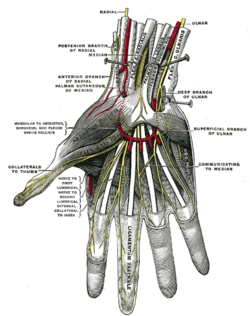 Superficial palmar nerves. (Deep branch of ulnar and superficial branch of ulnar labeled at center right.) | |
 Diagram of segmental distribution of the cutaneous nerves of the right upper extremity. (Ulnar palmar labeled at bottom left, in blue.) | |
| Details | |
| From | ulnar nerve |
| To | superficial branch, deep branch |
| Identifiers | |
| Latin | ramus palmaris nervi ulnaris |
| TA | A14.2.03.044 |
| FMA | 44878 |
The palmar branch of the ulnar nerve arises about five cm proximal to the wrist from where the ulnar nerve splits into palmar and dorsal branches.
The palmar branch represents the continuation of the ulnar nerve as it crosses the flexor retinaculum of the hand on the lateral side of the pisiform bone, medial to and a little behind the ulnar artery.
Some sources state that it ends by dividing into a superficial and a deep branch.[1] (Other sources state that the superficial branch of ulnar nerve and deep branch of ulnar nerve are the terminal branches of the ulnar nerve itself.)[2]
Additional Images
 Palmar branch of ulnar nerve
 Palmar branch of ulnar nerve
References
This article incorporates text in the public domain from the 20th edition of Gray's Anatomy (1918)
- ↑ The Anterior Divisions - Gray's Anatomy of the Human Body - Yahoo! Education
- ↑ Ellis, Harold; Susan Standring; Gray, Henry David (2005). Gray's anatomy: the anatomical basis of clinical practice. St. Louis, Mo: Elsevier Churchill Livingstone. p. 726. ISBN 0-443-07168-3.
External links
- Hand kinesiology at the University of Kansas Medical Center - palmar at left
This article is issued from Wikipedia - version of the 6/4/2016. The text is available under the Creative Commons Attribution/Share Alike but additional terms may apply for the media files.