Notochord
| Notochord | |
|---|---|
 Transverse section of a chick embryo of forty-five hours’ incubation. | |
| Details | |
| Precursor | chordamesoderm |
| Gives rise to | nucleus pulposus |
| Identifiers | |
| Latin | notochorda |
| MeSH | A16.254.610 |
| Code | TE E5.0.1.1.0.0.8 |
In animal anatomy, the notochord is a flexible rod made out of a material similar to cartilage. If a species has a notochord, it is, by definition, a chordate. The notochord lies along the anteroposterior ("head to tail") axis, is usually closer to the dorsal than the ventral surface of the animal, and is composed of cells derived from the mesoderm. The notochord has been observed to have many functions including developmental functions. The most commonly cited functions are as a site of muscle attachment, vertebral precursor, and as a midline tissue that provides signals to the surrounding tissue during development.[1]
Notochords are thought to be advantageous (both in an evolutionary and developmental context) because they provide(d) rigid structure for muscle attachment, but were still flexible. In some chordates, it persists throughout life as the main axial support of the body, while in most tetrapods it becomes the nucleus pulposus of the intervertebral disc.[1] The notochord plays a key role in signalling and coordinating development. Embryos of vertebrates still form transient notochord structures today during the gastrulation phase of development. The notochord is found ventral to the neural tube.
Development
Notogenesis is the development of the notochord by the epiblasts that make up the floor of the amnion cavity.[2] The notochord arises from the bilaminar embryonic disk. The notochord forms during gastrulation and soon after induces the formation of the neural plate (neurulation), synchronizing the development of the neural tube. On the ventral aspect of the neural groove an axial thickening of the endoderm takes place. (In bipedal chordates, e.g. humans, this surface is properly referred to as the anterior surface). This thickening appears as a furrow (the chordal furrow) the margins of which anastomose (come into contact), and so convert it into a solid rod of polygonal-shaped cells (the notochord) which is then separated from the endoderm.
It extends throughout the entire length of the future vertebral column, and reaches as far as the anterior end of the midbrain, where it ends in a hook-like extremity in the region of the future dorsum sellae of the sphenoid bone. Initially it exists between the neural tube and the endoderm of the yolk-sac, but soon becomes separated from them by the mesoderm, which grows medially and surrounds it. From the mesoderm surrounding the neural tube and notochord, the skull, vertebral column, and the membranes of the brain and medulla spinalis are developed.
A postembryonic vestige of the notochord is found in the nucleus pulposus of the intervertebral discs. Isolated notochordal remnants may escape their lineage-specific destination in the nucleus pulposus and instead attach to the outer surfaces of the vertebral bodies, from which notochordal cells largely regress.[3] In humans, by the age of 4, all notochord residue is replaced by a population of chondrocyte-like cells of unclear origin.[4] Persistence of notochordal cells within the vertebra may cause a pathologic condition: persistent notochordal canal.[5] They are also found to persist in the nasopharyngeal space and, in such an unusual instance, may give rise to Tornwaldt's cyst.
Neurology
Research into the notochord has played a key role in understanding the development of the central nervous system. By transplanting and expressing a second notochord near the dorsal neural tube, 180 degrees opposite of the normal notochord location, one can induce the formation of motor neurons in the dorsal tube. Motor neuron formation generally occurs in the ventral neural tube, while the dorsal tube generally forms sensory cells.
The notochord secretes a protein called sonic hedgehog homolog (SHH), a key morphogen regulating organogenesis and having a critical role in signaling the development of motor neurons.[6] The secretion of SHH by the notochord establishes the ventral pole of the dorsal-ventral axis in the developing embryo.
Evolution

The notochord is the defining feature of Chordates, and was present throughout life in many of the earliest chordates. Although the stomochord of hemichordates was once thought to be homologous, it is now viewed as a convergence.[7] Pikaia appears to have a proto-notochord, and notochords are present in several basal chordates such as Haikouella, Haikouichthys, and Myllokunmingia, all from the Cambrian. The Ordovician oceans included many diverse species of agnathan fish which possessed notochords, either with attached bony elements or without, most notably the conodonts,[8] placoderms[9] and ostracoderms. Even after the evolution of the vertebral column in chondrichthyes and osteichthyes, these taxa remained common and are well represented in the fossils record. Several species (see list below) have reverted to the primitive state, retaining the notochord into adulthood, though the reasons for this are not well-understood. Scenarios for the evolutionary origin of the notochord have been comprehensively reviewed (Annona, G., Holland, N. D., and D'Aniello, S. 2015. Evolution of the notochord. EvoDevo 6: article 30). They point out that, although many of these ideas have not been well supported by advances in molecular phylogenetics and developmental genetics, two of them have actually been revived under the stimulus of modern molecular approaches(the first proposes that the notochord evolved de novo in chordates, and the second derives it from a homologous structure, the axochord, that was present in annelid-like ancestors of the chordates). Deciding between these two scenarios (or possibly another yet to be proposed) should be facilitated by much more thorough studies of gene regulatory networks in a wide spectrum of animals.
Structure
The notochord is composed primarily of a core of glycoproteins, encased in a sheath of collagen fibers wound into two opposing helices. The angle between these fibers determines whether increased pressure in the core will result in shortening and thickening versus lengthening and thinning.[10]
Organisms which retain a post-embryonic notochord
- Amphioxus
- Tunicate larvae
- Hagfish
- Lamprey
- Sturgeon
- Coelacanth
- African lungfish
- Tadpoles
- Ostracoderms (extinct)
Additional images
 Surface view of embryo of Concolor gibbon (Hylobates concolor).
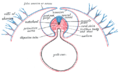 Diagram of a transverse section, showing the mode of formation of the amnion in the chick.
 Section through the head of a human embryo, about twelve days old, in the region of the hind-brain.
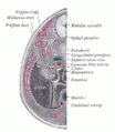 Transverse section of human embryo eight and a half to nine weeks old.
References
- 1 2 Stemple, Derek L. (2005-06-01). "Structure and function of the notochord: an essential organ for chordate". Development. 132 (11): 2503–2512. doi:10.1242/dev.01812. ISSN 0950-1991. PMID 15890825.
- ↑ The trilaminar germ disk (3rd week)
- ↑ Choi, K.; Cohn, Martin J.; Harfe, Brian D. (2009). "Identification of Nucleus Pulposus Precursor Cells and Notochordal Remnants in the Mouse: Implications for Disk Degeneration and Chordoma Formation". Developmental Dynamics. 237 (12): 3953–3958. doi:10.1002/dvdy.21805. PMC 2646501
 . PMID 19035356.
 - ↑ Urban, J. P. G. (2000). "The Nucleus of the Intervertebral Disc from Development to Degeneration". Integrative and Comparative Biology. 40: 53. doi:10.1093/icb/40.1.53.
- ↑ Christopherson, Lr; Rabin, Bm; Hallam, Dk; Russell, Ej (1 January 1999). "Persistence of the notochordal canal: MR and plain film appearance" (Free full text). AJNR. American journal of neuroradiology. 20 (1): 33–6. ISSN 0195-6108. PMID 9974055.
- ↑ Echelard, Y; Epstein, Dj; St-Jacques, B; Shen, L; Mohler, J; Mcmahon, Ja; Mcmahon, Ap (December 1993). "Sonic hedgehog, a member of a family of putative signaling molecules, is implicated in the regulation of CNS polarity". Cell. 75 (7): 1417–30. doi:10.1016/0092-8674(93)90627-3. PMID 7916661.
- ↑ Kardong, Kenneth V. (1995). Vertebrates: comparative anatomy, function, evolution. McGraw-Hill. pp. 55, 57. ISBN 0-697-21991-7.
- ↑ http://www.palaeos.com/Vertebrates/Units/030Conodonta/030.000.html
- ↑ http://www.palaeos.com/Vertebrates/Units/Unit060/060.000.html
- ↑ M. A. R. Koehl. "Mechanical Design of Fiber-Wound Hydraulic Skeletons: The Stiffening and Straightening of Embryonic Notochords".