Neurogastroenterology
Neurogastroenterology encompasses the study of the brain, the gut, and their interactions with relevance to the understanding and management of gastrointestinal motility and functional gastrointestinal disorders. Specifically, neurogastroenterology focuses on the functions, malfunctions, and the malformations of the sympathetic, parasympathetic, and enteric divisions of the digestive tract.[1]
Function of neurons in the gastrointestinal tract
The peristaltic reflex
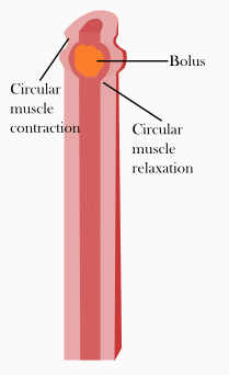
Peristalsis is a series of radially symmetrical contractions and relaxations of muscles which propagate down a muscular tube. In humans and other mammals, peristalsis is found in the smooth muscles of the digestive tract to propel contents through the digestive system. The word is derived from New Latin and comes from the Greek peristallein, "to wrap around," from peri-, "around" + stallein, "to place". Peristalsis was discovered in 1899 by the work of physiologists William Bayliss and Ernest Starling. Working on the small intestines of dogs, they found that the response of increasing the pressure in the intestine caused the contraction of the muscle wall above the point of stimulation and the relaxation of the muscle wall below the point of stimulation.[2][3]
Segmentation
Segmentation contractions are the contractions in intestines carried out by the smooth muscle walls. Unlike peristalsis, which involves the contraction and relaxation of muscles in one direction, segmentation occurs simultaneously in both directions as the circular muscles alternatively contract. This allows for thorough mixing of intestinal contents, known as chyme, to allow greater absorption.
Secretion
The secretion of gastrointestinal enzymes, such as gastrin and secretin, is regulated through cholinergic neurons residing in the walls of the digestive tract. Hormone secretion is controlled by the vagovagal reflex, where the neurons in the digestive tract communicate through both afferent and efferent pathways with the vagus nerve.[4]
Anatomy
Enteric nervous system
The enteric nervous system is one of the main divisions of the nervous system and is the main focus of neurogastroenterology. The enteric nervous system refers to the entire system of neurons that govern the gastrointestinal system.[5] It is capable of operating independently of the brain and spinal cord,[3] but does rely on innervation from the autonomic nervous system via the vagus nerve and prevertebral ganglia in healthy subjects. However, studies have shown that the system is operable with a severed vagus nerve.[6] The neurons of the enteric nervous system control the motor functions of the system, in addition to the secretion of gastrointestinal enzymes. These neurons communicate through many neurotransmitters similar to the CNS, including acetylcholine, dopamine, and serotonin. The large presence of serotonin and dopamine in the gut are key areas of research for neurogastroenterologists.[7][8][9]
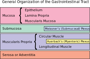
Auerbach's plexus
Auerbach's plexus, also known as the myenteric plexus, is a collection of unmyelinated fibers and postganglionic autonomic cell bodies that lie between the circular and longitudinal layers of the muscularis externa in the gastrointestinal tract. It was discovered and named by German neuropathologist Leopold Auerbach. These neurons provide motor inputs to both layers of the muscularis externa, and provide both parasympathetic and sympathetic input. The anatomy of the plexus is similar to the anatomy of the central nervous system. The plexus includes sensory receptors, such as chemoreceptors and mechanoreceptors, that are used to provide sensory input to the interneurons in the enteric nervous system. The plexus is the parasympathetic nucleus of origin for the vagus nerve, and communicate with the medulla oblongata through both the anterior and posterior vagal nerves.
Meissner's plexus
Meissner's plexus is a collection of the plexuses of parasympathetic nerves that run from Auerbach's plexus to the muscularis mucosae of the gastrointestinal wall. It was discovered and named by German physiologist Georg Meissner. It functions as a pathway for the innervation in the mucosa layer of the gastrointestinal wall.
Disorders
Functional gastrointestinal disorders
Functional gastrointestinal (GI) disorders are a class of gastrointestinal disorders where there is a malfunction in the normal activities of the gastrointestinal tract, but there are no structural abnormalities that can explain the cause. There are rarely any tests that can detect the presence of these disorders. Clinical research in neurogastroenterology focuses mainly on the study of common functional gastrointestinal disorders such as irritable bowel syndrome, the most common functional GI disorder.[10]
Motility disorders
Motility disorders are the second classification of gastrointestinal disorder studied by neurogastroenterologists. Motility disorders are divided by what they affect, with four regions: The esophagus, the stomach, the small intestines, and the large intestines. Clinical research in neurogastroenterology focuses mainly on the study of common motility disorders such as gastroesophageal reflux disease, the damage of the mucosa of the esophagus caused by rising stomach acid through the lower esophageal sphincter.[11]
Neurogastroenterology societies
- American Neurogastroenterology and Motility Society[12]
- European Society of Neurogastroenterology and Motility[13]
See also
Additional images
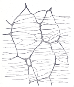 The myenteric plexus of a rabbit. X 50.
 The submucosal plexus of a rabbit. X 50.
References
- ↑ Wood, JD; DH Alpers; PLR Andrews (1999). "Fundamentals of Neurogastroenterology". Gut. 45: 6–16. doi:10.1136/gut.45.2008.ii6.
- ↑ Keet, A. D. "The Pyloric Sphincteric Cylinder in health and disease". Retrieved 18 November 2013.
- 1 2 Gershon, Michael (1998). The Second Brain. New York: HarperCollins. pp. 2–7. ISBN 0-06-018252-0.
- ↑ Herman MA, Cruz MT, Sahibzada N, Verbalis J, Gillis RA (January 2009). "GABA signaling in the nucleus tractus solitarius sets the level of activity in dorsal motor nucleus of the vagus cholinergic neurons in the vagovagal circuit". Am. J. Physiol. Gastrointest. Liver Physiol. 296 (1): G101–11. doi:10.1152/ajpgi.90504.2008. PMC 2636929
 . PMID 19008339.
 - ↑ John Barton Furness (15 April 2008). The Enteric Nervous System. John Wiley & Sons. ISBN 978-1-4051-7344-5. Retrieved 16 January 2013.
- ↑ Li,Ying; Owyang,Chung (September 2003). "Musings on the Wanderer: What's New in Our Understanding of Vago-Vagal Reflexes? V. Remodeling of vagus and enteric neural circuitry after vagal injury". American Journal of Physiology. Gastrointestinal and Liver Physiology. 285 (3): G461–9. doi:10.1152/ajpgi.00119.2003.
- ↑ Pasricha, Pankaj Jay. "Stanford Hospital: Brain in the Gut - Your Health".
- ↑ Martinucci I et al. Genetics and pharmacogenetics of aminergic transmitter pathways in functional gastrointestinal disorders. Pharmacogenomics. 2015;16(5):523-39. Review. PMID 25916523
- ↑ Smitka K, et al. The role of "mixed" orexigenic and anorexigenic signals and autoantibodies reacting with appetite-regulating neuropeptides and peptides of the adipose tissue-gut-brain axis: relevance to food intake and nutritional status in patients with anorexia nervosa and bulimia nervosa. Int J Endocrinol. 2013;2013:483145. Review. PMID 24106499 Free full text PMC 3782835
- ↑ Kumar, A.; Rinwa P.; Sharma N. (2012). "Irritable Bowel Syndrome: A Review". J Phys Pharm Adv. 2 (2): 97–108.
- ↑ DeVault KR, Castell DO (1999). "Updated guidelines for the diagnosis and treatment of gastroesophageal reflux disease. The Practice Parameters Committee of the American College of Gastroenterology". Am J Gastroenterol. 94 (6): 1434–42. doi:10.1111/j.1572-0241.1999.1123_a.x. PMID 10364004.
- ↑ ANMS - American Neurogastroenterology and Motility Society
- ↑ ESNM - European Society for Neurogastroenterology & Motility