Histology

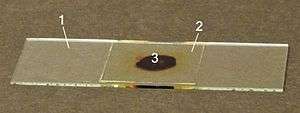
- glass microscope slide
- glass coverslip
- stained tissue section, mounted between 1. and 2.
Histology[help 1] is the study of the microscopic anatomy (microanatomy) of cells and tissues of plants and animals. It is commonly performed by examining cells and tissues under a light microscope or electron microscope, the specimen having been sectioned (cut into a thin cross section with a microtome), stained, and mounted on a microscope slide. Histological studies may be conducted using tissue culture, where live human or animal cells are isolated and maintained in an artificial environment for various research projects. The ability to visualize or differentially identify microscopic structures is frequently enhanced through the use of histological stains. Histology is an essential tool of biology and medicine.
Histopathology, the microscopic study of diseased tissue, is an important tool in anatomical pathology, since accurate diagnosis of cancer and other diseases usually requires histopathological examination of samples. Trained physicians, frequently licensed pathologists, are the personnel who perform histopathological examination and provide diagnostic information based on their observations. The trained personnel who prepare histological specimens for examination are histotechnicians, histology technicians (HT), histology technologists (HTL), medical scientists, medical laboratory technicians, or biomedical scientists. Their field of study is called histotechnology.
Sample preparation
Fixing
Chemical fixation with formaldehyde or other chemicals
Chemical fixatives are used to preserve tissue from degradation, and to maintain the structure of the cell and of sub-cellular components such as cell organelles (e.g., nucleus, endoplasmic reticulum, mitochondria). The most common fixative for light microscopy is 10% neutral buffered formalin (4% formaldehyde in phosphate buffered saline). For electron microscopy, the most commonly used fixative is glutaraldehyde, usually as a 2.5% solution in phosphate buffered saline. These fixatives preserve tissues or cells mainly by irreversibly cross-linking proteins. The main action of these aldehyde fixatives is to cross-link amino groups in proteins through the formation of methylene bridges (-CH2-), in the case of formaldehyde, or by C5H10 cross-links in the case of glutaraldehyde. This process, while preserving the structural integrity of the cells and tissue can damage the biological functionality of proteins, particularly enzymes, and can also denature them to a certain extent. This can be detrimental to certain histological techniques. Further fixatives are often used for electron microscopy such as osmium tetroxide or uranyl acetate
Formalin fixation leads to degradation of mRNA, miRNA and DNA in tissues. However, extraction, amplification and analysis of these nucleic acids from formalin-fixed, paraffin-embedded tissues is possible using appropriate protocols.[1]
Frozen section fixation
Frozen section procedure is a rapid way to fix and mount histology sections using a refrigeration device called a cryostat. It is often used after surgical removal of tumors to allow rapid determination of margin (that the tumor has been completely removed).
Processing - dehydration, clearing, and infiltration
The aim of Tissue Processing is to remove water from tissues and replace with a medium that solidifies to allow thin sections to be cut. Biological tissue must be supported in a hard matrix to allow sufficiently thin sections to be cut, typically 5 μm (micrometres; 1000 micrometres = 1 mm) thick for light microscopy and 80-100 nm (nanometre; 1,000,000 nanometres = 1 mm) thick for electron microscopy. For light microscopy, paraffin wax is most frequently used. Since it is immiscible with water, the main constituent of biological tissue, water must first be removed in the process of dehydration. Samples are transferred through baths of progressively more concentrated ethanol to remove the water. This is followed by a hydrophobic clearing agent (such as xylene) to remove the alcohol, and finally molten paraffin wax, the infiltration agent, which replaces the xylene. Paraffin wax does not provide a sufficiently hard matrix for cutting very thin sections for electron microscopy. Instead, resins are used. Epoxy resins are the most commonly employed embedding media, but acrylic resins are also used, particularly where immunohistochemistry is required. Thicker sections (0.35μm to 5μm) of resin-embedded tissue can also be cut for light microscopy. Again, the immiscibility of most epoxy and acrylic resins with water necessitates the use of dehydration, usually with ethanol.
Embedding
.png)
After the tissues have been dehydrated, cleared, and infiltrated with the embedding material, they are ready for external embedding. During this process the tissue samples are placed into molds along with liquid embedding material (such as agar, gelatine, or wax) which is then hardened. This is achieved by cooling in the case of paraffin wax and heating (curing) in the case of the epoxy resins. The acrylic resins are polymerised by heat, ultraviolet light, or chemical catalysts. The hardened blocks containing the tissue samples are then ready to be sectioned.
Because Formalin-fixed, paraffin-embedded (FFPE) tissues may be stored indefinitely at room temperature, and nucleic acids (both DNA and RNA) may be recovered from them decades after fixation, FFPE tissues are an important resource for historical studies in medicine.
Embedding can also be accomplished using frozen, non-fixed tissue in a water-based medium. Pre-frozen tissues are placed into molds with the liquid embedding material, usually a water-based glycol, OCT, TBS, Cryogel, or resin, which is then frozen to form hardened blocks.
Sectioning
For light microscopy, a steel knife mounted in a microtome is used to cut 4-micrometer-thick tissue sections which are mounted on a glass microscope slide. For transmission electron microscopy, a diamond knife mounted in an ultramicrotome is used to cut 50-nanometer-thick tissue sections which are mounted on a 3-millimeter-diameter copper grid. Then the mounted sections are treated with the appropriate stain.
Sections can be cut through the tissue in a number of directions. For pathological evaluation of tissues, vertical sectioning, (cut perpendicular to the surface of the tissue to produce a cross section) is the usual method. Horizontal (also known as transverse or longitudinal) sectioning, cut along the long axis of the tissue, is often used in the evaluation of the hair follicles and pilosebaceous units. Tangential to horizontal sectioning is used in Mohs surgery and in methods of CCPDMA.
Cryosectioning
Fixed or unfixed tissue may be frozen and sliced using a microtome mounted in a refrigeration device known as a cryostat. The frozen sections are mounted on a glass slide and may be stained to enhance the contrast between different tissues. Unfixed frozen sections can also be used for studies requiring enzyme localization in tissues and cells. It is necessary to fix tissue for certain procedures such as antibody linked immunofluorescence staining. Frozen sectioning can also be used to determine if a tumour is malignant when it is found incidentally during surgery on a patient.

Staining
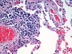
Biological tissue has little inherent contrast in either the light or electron microscope. Staining is employed to give both contrast to the tissue as well as highlighting particular features of interest. Where the underlying mechanistic chemistry of staining is understood, the term histochemistry is used. Hematoxylin and eosin (H&E stain) is the most commonly used light microscopical stain in histology and histopathology. Hematoxylin, a basic dye, stains nuclei blue due to an affinity to nucleic acids in the cell nucleus; eosin, an acidic dye, stains the cytoplasm pink. Uranyl acetate and lead citrate are commonly used to impart contrast to tissue in the electron microscope.
There are many other staining techniques that have been used to selectively stain cells and cellular components. One of these techniques involves marking peripheral tumors or surgical margins, in which a certain color of dye is applied to the posterior border of a sample, another to the anterior, etc., so that one can identify the location of a tumor or other pathology within a specimen. Other compounds used to color tissue sections include safranin, Oil Red O, Congo red, Fast green FCF, silver salts, and numerous natural and artificial dyes that usually originated from the development of dyes for the textile industry.
Histochemistry refers to the science of using chemical reactions between laboratory chemicals and components within tissue. A commonly performed histochemical technique is the Perls Prussian blue reaction, used to demonstrate iron deposits in diseases like hemochromatosis.
Histology samples have often been examined by radioactive techniques. In historadiography, a slide (sometimes stained histochemically) is X-rayed. More commonly, autoradiography is used to visualize the locations to which a radioactive substance has been transported within the body, such as cells in S phase (undergoing DNA replication) which incorporate tritiated thymidine, or sites to which radiolabeled nucleic acid probes bind in in situ hybridization. For autoradiography on a microscopic level, the slide is typically dipped into liquid nuclear tract emulsion, which dries to form the exposure film. Individual silver grains in the film are visualized with dark field microscopy.
Recently, antibodies have been used to specifically visualize proteins, carbohydrates, and lipids. This process is called immunohistochemistry, or when the stain is a fluorescent molecule, immunofluorescence. This technique has greatly increased the ability to identify categories of cells under a microscope. Other advanced techniques, such as nonradioactive in situ hybridization, can be combined with immunochemistry to identify specific DNA or RNA molecules with fluorescent probes or tags that can be used for immunofluorescence and enzyme-linked fluorescence amplification (especially alkaline phosphatase and tyramide signal amplification). Fluorescence microscopy and confocal microscopy are used to detect fluorescent signals with good intracellular detail. Digital cameras are increasingly used to capture histological and histopathological image
Common laboratory stains
| Stain | Common use | Nucleus | Cytoplasms | Red blood cell (RBC) | Collagen fibers | Specifically stains |
|---|---|---|---|---|---|---|
| Haematoxylin | General staining when paired with eosin (i.e. H&E) | Orange, Cyan Blue or Green | Blue/Brown/Black | N/A | N/A | Nucleic acids—blue ER (endoplasmic reticulum)—blue |
| Eosin | General staining when paired with haematoxylin (i.e. H&E) | N/A | Pink | Orange/red | Pink | Elastic fibers—pink Collagen fibers—pink Reticular fibers—pink |
| Toluidine blue | General staining | Blue | Blue | Blue | Blue | Mast cells granules—purple |
| Masson's trichrome stain | Connective tissue | Black | Red/pink | Red | Blue/green | Cartilage—blue/green Muscle fibers—red |
| Mallory's trichrome stain | Connective tissue | Red | Pale red | Orange | Deep blue | Keratin—orange
Cartilage—blue |
| Weigert's elastic stain | Elastic fibers | Blue/black | N/A | N/A | N/A | Elastic fibers—blue/black |
| Heidenhain's AZAN trichrome stain | Distinguishing cells from extracellular components | Red/purple | Pink | Red | Blue | Muscle fibers—red Cartilage—blue Bone matrix—blue |
| Silver stain | Reticular fibers, nerve fibers, fungi | N/A | N/A | N/A | N/A | Reticular fibers—brown/black Nerve fibers—brown/black Fungi—black |
| Wright's stain | Blood cells | Bluish/purple | Bluish/gray | Red/pink | N/A | Neutrophil granules—purple/pink Eosinophil granules—bright red/orange Basophil granules—deep purple/violet Platelet granules—red/purple |
| Orcein stain | Elastic fibres | Deep blue | N/A | Bright red | Pink | Elastic fibres—dark brown Mast cells granules—purple Smooth muscle—light blue |
| Periodic acid-Schiff stain (PAS) | Basement membrane, localizing carbohydrates | Blue | N/A | N/A | Pink | Glycogen and other carbohydrates—magenta |
Table sourced from Michael H. Ross; Wojciech Pawlina (2006). Histology: A Text and Atlas. Hagerstown, MD: Lippincott Williams & Wilkins. ISBN 0-7817-5056-3.
The Nissl method and Golgi's method are useful in identifying neurons.
Alternative techniques
Plastic embedding is commonly used in the preparation of material for electron microscopy. Tissues are embedded in epoxy resin. Very thin sections (less than 0.1 micrometer) are cut using diamond or glass knives. The sections are stained with electron dense stains (uranium and lead) so that they can be seen with the electron microscope.
History

In the 17 century, Italian Marcello Malpighi invented one of the first microscopes for studying tiny biological entities. Malpighi analysed several parts of the organs of bats, frogs and other animals under the microscope. Malpighi, while studying the structure of the lung, noticed its membranous alveoli and the hair-like connections between veins and arteries, which he named capillaries. His discovery established how the oxygen we breathe enters the blood stream and serves the body.[2]
In the 19th century, histology was an academic discipline in its own right. The 1906 Nobel Prize in Physiology or Medicine was awarded to histologists Camillo Golgi and Santiago Ramon y Cajal. They had dueling interpretations of the neural structure of the brain based in differing interpretations of the same images. Cajal won the prize for his correct theory and Golgi for the staining technique he invented to make it possible.
Histological classification of animal tissues
There are four basic types of tissues: muscle tissue, nervous tissue, connective tissue, and epithelial tissue. All tissue types are subtypes of these four basic tissue types (for example, blood is classified as connective tissue, since the blood cells are suspended in an extracellular matrix, the plasma).
- Epithelium: the lining of glands, bowel, skin, and some organs like the liver, lung, and kidney
- Endothelium: the lining of blood and lymphatic vessels
- Mesothelium: the lining of pleural and pericardial spaces
- Mesenchyme: the cells filling the spaces between the organs, including fat, muscle, bone, cartilage, and tendon cells
- Blood cells: the red and white blood cells, including those found in lymph nodes and spleen
- Neurons: any of the conducting cells of the nervous system
- Germ cells: reproductive cells (spermatozoa in men, oocytes in women)
- Placenta: an organ characteristic of true mammals during pregnancy, joining mother and offspring, providing endocrine secretion and selective exchange of soluble, but not particulate, blood-borne substances through an apposition of uterine and trophoblastic vascularised parts
- Stem cells: cells with the ability to develop into different cell types
Note that tissues from plants, fungi, and microorganisms can also be examined histologically. Their structure is very different from animal tissues.
Related sciences
- Cell biology is the study of living cells, their DNA and RNA and the proteins they express.
- Anatomy is the study of organs visible by the naked eye.
- Morphology studies entire organisms.
- Cytology is the microscopic study of loose cells or clusters obtained from bodily secretions, aspirations, scrapes, swipes, or washings.
Artifacts
Artifacts are structures or features in tissue that interfere with normal histological examination. These are not always present in normal tissue and can come from outside sources. Artifacts interfere with histology by changing the tissues appearance and hiding structures. These can be divided into two categories:
Pre-histology
These are features and structures that have been introduced prior to the collection of the tissues. A common example of these include: ink from tattoos and freckles (melanin) in skin samples.
Post-histology
Artifacts can result from tissue processing. Processing commonly leads to changes like shrinkage, washing out of particular cellular components, color changes in different tissues types and alterations of the structures in the tissue. Because these are caused in a laboratory the majority of post histology artifacts can be avoided or removed after being discovered. A common example is mercury pigment left behind after using Zenker's fixative to fix a section.
Histology art
Normal patterning of tissues and artifacts resulting from the tissue preparation process ensure that each histological section is unique. Like a piece of biological art these images provide a deep insight into the organization and function of our bodies. Histological patterns that look like everyday objects or features are emerging on social and scientific communities [3] and even in histopathology journal articles.[4] Histology is an area of science where art and science collide. It demonstrates that histology can be appreciated by not only the detail-oriented pathologist but also the art loving layperson and is making histology and pathology more accessible and less daunting as a complex science.
See also
- Tissue
- Anatomical pathology
- Automated tissue image analysis
- Biological staining
- Geoffrey Bourne
- Gross anatomy
- Cooperative Human Tissue Network (CHTN)
- Digital Pathology
- Arthur Worth Ham
- Histopathology
- Important publications in histology (Arthur Worth Ham and David H. Cormack's Histology, for example)
- Laser capture microdissection
- Pathology
- Cytoarchitecture
- Plant anatomy
- Extracellular matrix
- Cell adhesion molecule
- Multicellularity
Notes
References
- ↑ Weiss AT, Delcour NM, Meyer A, Klopfleisch R (2010). "Efficient and Cost-Effective Extraction of Genomic DNA From Formalin-Fixed and Paraffin-Embedded Tissues". Veterinary Pathology. 227 (4): 834–8. doi:10.1177/0300985810380399. PMID 20817894.
- ↑ Adelmann, Howard (1966) Marcello Malpighi and the Evolution of Embryology 5 vol., Cornell University Press, Ithaca, N.Y. OCLC 306783
- ↑ i-heart-histo. "Histological art".
- ↑ Coyne J. (2012). "A squamous cell carcinoma with a Saint Valentine's day message". Int J Surg Pathol. 20 (1): 62. doi:10.1177/1066896911434768. PMID 22287650.
- Merck Source (2002). Dorland's Medical Dictionary. Retrieved 2005-01-26.
- Stedman's Medical Dictionaries (2005). Stedman's Online Medical Dictionary. Retrieved 2005-01-26.
- 4,000 online histology images (2007). <http://histology-online.com>
External links
| Wikimedia Commons has media related to Histology. |
| Library resources about Histology |
- HistoNet 2000 - Online Interactive Atlas
- Histology Protocols
- Histoweb
- SIU SOM Histology
- Visual Histology Atlas
- Histology Glossary
- Histology Group of Victoria Incorporated
- Histology Photomicrographs
- Histology-online
- Virtual Slidebox
- Blue Histology
- Dr. Kasem Histology Homepage
- Histology atlas and more
- Medical Histology Lecture Series by Dr. Krause, Department of Pathology and Anatomical Science, University of Missouri School of Medicine