Lobomycosis
| Lobo's disease | |
|---|---|
 | |
| Histopathological changes in the skin seen in lobomycosis. Source: CDC. | |
| Classification and external resources | |
| Specialty | infectious disease |
| ICD-10 | B48.0 |
| ICD-9-CM | 116.2 |
| DiseasesDB | 32590 |
| eMedicine | derm/832 |

Lobomycosis[1] also known as (Jorge) Lobo's disease or lacaziosis,[2] is a blastomycosis, a fungal infection of the skin caused by Lacazia loboi (formerly named Loboa loboi),[3] and discovered by Brazilian dermatologist Jorge Lobo. Other names which were given to the disease are: keloidal blastomycosis, Amazonian blastomycosis, blastomycoid granuloma, miraip and piraip. These last two names were given by natives of the Amazon and mean that which burns.
This disease is usually found in humans[4] and bottle-nosed dolphins.
Clinical features
The disease is endemic in rural regions in South America and Central America.
Infection most commonly develops after minor scratches or insect bites, but many patients cannot recall any skin trauma. Human-to-human transmission does not occur, and the disease is only acquired from the environment.[5] The appearances are of a chronic keloidal nodular lesions occur on the face, ears, or extremities.
Diagnosis of Lobo's disease is made by taking a sample of the infected skin (a skin biopsy) and examining it under the microscope. Lacazia loboi is characterized by long chains of spherical cells interconnected by tubules. The cells appear to be yeast-like with a diameter of 5 to 12 μm. Attempts to culture L. loboi have so far been unsuccessful.
Differential diagnosis
The disease is often misdiagnosed as Blastomyces dermatitidis or Paracoccidiodes brasiliensis due to its similar morphology.
Treatment
Surgical excision or cryosurgery is the treatment of choice.[6] Treatment with antifungals has been considered ineffective, but the use of clofazimine and dapsone in patients with leprosy and lobomycosis has been found to improve the latter. This treatment regimen, with concomitant itraconazole, has been used to prevent recurrence after surgery.[7]
Lobo's disease in dolphins
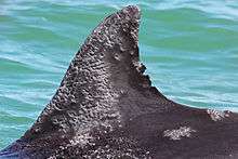
Lesions in dolphins occur on the dorsal fin, head, flukes, and peduncle. In January 2006, a potential epidemic of lobomycosis was reported in dolphins of the Indian River Lagoon in Florida.[8]
See also
References
- ↑ Talhari C, Oliveira CB, de Souza Santos MN, Ferreira LC, Talhari S (June 2008). "Disseminated lobomycosis". Int. J. Dermatol. 47 (6): 582–3. doi:10.1111/j.1365-4632.2008.03678.x. PMID 18477148.
- ↑ Xavier MB, Libonati RM, Unger D, et al. (February 2008). "Macrophage and TGF-beta immunohistochemical expression in Jorge Lobo's disease". Hum. Pathol. 39 (2): 269–74. doi:10.1016/j.humpath.2007.06.016. PMID 17959227.
- ↑ Honda, Kord; Horner, Kyle (2006). "Lobomycosis". eMedicine. Retrieved 2007-01-18.
- ↑ Elsayed S, Kuhn SM, Barber D, Church DL, Adams S, Kasper R (April 2004). "Human case of lobomycosis". Emerging Infect. Dis. 10 (4): 715–8. doi:10.3201/eid1004.030416. PMC 3323076
 . PMID 15200867.
 - ↑ Baruzzi RG, Lacaz CS, Souza FA (1979). "História natural da doença de Jorge Lobo. Ocorrência entre os índios Caibi (Brasil Central)". Rev Inst Med Trop Sao Paulo. 21: 302–338.
- ↑ Sobera JO, Elewski BE (2007). "Fungal diseases". In Bolognia JL. Dermatology. St. Louis: Mosby. p. 1150. ISBN 1-4160-2999-0.
- ↑ Rosa PS, Soares CT, Belone AF, et al. (2009). "Accidental Jorge Lobo's disease in a worker dealing with Lacazia loboi infected mice: a case report". Journal of Medical Case Reports. 3: 67. doi:10.1186/1752-1947-3-67.
- ↑ Reif JS, Mazzoil MS, McCulloch SD, et al. (January 2006). "Lobomycosis in Atlantic bottlenose dolphins from the Indian River Lagoon, Florida". J Am Vet Med Assoc. 228 (1): 104–8. doi:10.2460/javma.228.1.104. PMID 16426180.
Further reading
- Rodríguez-Toro G (May 1993). "Lobomycosis". Int. J. Dermatol. (Review). 32 (5): 324–32. doi:10.1111/j.1365-4362.1993.tb01466.x. PMID 8505156.