Levator scapulae muscle
| Levator scapulae muscle | |
|---|---|
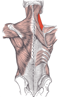 Muscles connecting the upper extremity to the vertebral column. (Levator scapula visible at upper right, at the neck.) | |
 Muscles of neck. (Levator scapula visible at center left.) | |
| Details | |
| Origin | Posterior tubercles of transverse processes of C1 - C4 vertebrae |
| Insertion | Superior part of medial border of scapula |
| Artery | dorsal scapular artery |
| Nerve | cervical nerve (C3, C4) and dorsal scapular nerve (C5) |
| Actions | Elevates scapula and tilts its glenoid cavity inferiorly by rotating scapula |
| Identifiers | |
| Latin | musculus levator scapulae |
| TA | A04.3.01.009 |
| FMA | 32519 |
In human anatomy, the levator scapulae (/ˌlᵻˈveɪtər ˈskæpjᵿliː/) is a skeletal muscle situated at the back and side of the neck. As the Latin name suggests, its main function is to lift the scapula.
Human anatomy
Origin and insertion
The levator scapulae originates from the posterior tubercle of the transverse process of cervical vertebrae one to four. The muscle is inserted into medial border of the scapula extending from superior angle to junction of spine and medial border of scapula [1]
The levator scapulae may lie deep to the sternocleidomastoideus at its origin, deep or adjacent to the splenius capitis at its origin and mid-portion, and deep to the trapezius in its lower portion.
Actions
When the spine is fixed, levator scapulae elevates the scapula and rotates its inferior angle medially.[1] It often works in combination with other muscles like the rhomboids and pectoralis minor to rotate down.
Elevating or rotating one shoulder at a time would require muscles to stabilize the cervical spine and keep it immobile so it does not flex or rotate. Elevating both at once with equal amounts of pull on both side of cervical spinal origins would counteract these forces. Downward rotation would be prevented by co-contraction of other muscles that elevate the spine, the upper fibers of the trapezius, which is an upward rotator.
When the shoulder is fixed, levator scapula rotates and flexes the cervical spine laterally.[2] When both shoulders are fixed, a simultaneous co-contraction of both levator scapulae muscles in equal amounts would not produce lateral flexion or rotation, and may produce straight flexion or extension of the cervical spine.
Relations
One of the muscles within the floor of the posterior triangle of the neck, the superior part of levator scapula is covered by sternocleidomastoid and its inferior part by the trapezius. [3] It is bounded in front by the scalenus medius and behind by splenius cervicis. The spinal accessory nerve crosses laterally in the middle part of the muscle and the dorsal scapular nerve may lie deep to or pass through it. [4]
Variations
The number of vertebral attachments varies; a slip may extend to the occipital or mastoid, to the trapezius, scalene or serratus anterior, or to the first or second rib. The muscle may be subdivided into several distinct parts from origin to insertion. Levator claviculæ from the transverse processes of one or two upper cervical vertebræ to the outer end of the clavicle corresponds to a muscle of lower animals. More or less union with the serratus anterior. [5]
Innervation and blood supply
The levator scapulae is supplied by two or three branches of the fourth and fifth cervical nerves,[1] and frequently by a branch from the dorsal scapular.[5]
The levator scapulae is supplied by the dorsal scapular artery. Normally, this artery has a small branch which passes laterally to the supraspinatus fossa of the scapula, and in a third of cases, this branch supplies the muscle. If the dorsal scapular artery comes off the transverse cervical artery, the parent transverse cervical artery splits, the dorsal scapular artery passes medially, while the transverse cervical artery passes laterally. [4]
Evolutionary variation
The muscles of the shoulder can be categorized into three topographic units: the scapulohumeral, axiohumeral, and axioscapular groups. Levator scapulae forms part of the latter group together with rhomboid major, rhomboid minor, serratus anterior, and trapezius. The trapezius evolved separately, but the other three muscles in this group evolved from the first eight or ten ribs and the transverse processes of the cervical vertebrae (homologous to the ribs). The serratus anterior formed the basal unit for these three muscles. In higher primates it has evolved into two separate muscles — serratus anterior and levator scapulae — by concentration of the proximal and distal fibers and progressive reduction of the intermediate fibers. The fibers concerned with the cranial displacement of the scapula became the levator scapulae. [6]
Additional images
 Position of levator scapulae muscle.
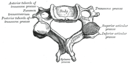 A cervical vertebra
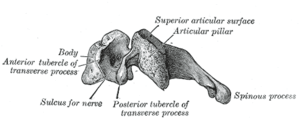 Side view of a typical cervical vertebra
 Left scapula. Dorsal surface.
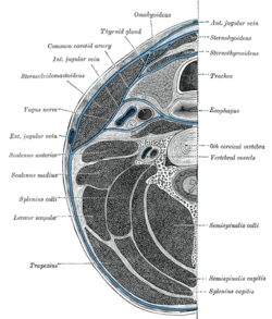 Section of the neck at about the level of the sixth cervical vertebra.
 Muscles of the neck. Lateral view.
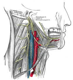 Hypoglossal nerve, cervical plexus, and their branches.
 The right brachial plexus with its short branches, viewed from in front.
 Levator scapulae muscle
 Brachial plexus. Deep dissection.
 Brachial plexus. Deep dissection. Anterolateral view
See also
| Wikimedia Commons has media related to Levator scapulae muscles. |
- Levator claviculae muscle
- Stiff neck - most commonly caused by pain in the Levator scapulae
Notes
This article incorporates text in the public domain from the 20th edition of Gray's Anatomy (1918)
- 1 2 3 Platzer 2004, p. 144
- ↑ Muscles/LevatorScapulae at exrx.net
- ↑ -154140595 at GPnotebook
- 1 2 Rockwood et al. 2009, p. 55
- 1 2 Gray's Anatomy (1918), see infobox
- ↑ Brand 2008, pp. 540–41
References
- Brand, R. A. (2008). "Origin and Comparative Anatomy of the Pectoral Limb". Clinical Orthopaedics and Related Research. 466 (3): 531–42. doi:10.1007/s11999-007-0102-6. PMC 2505211
 . PMID 18264841.
 - Platzer, Werner (2004). Color Atlas of Human Anatomy, Vol. 1: Locomotor System (5th ed.). Thieme. ISBN 3-13-533305-1.
- Rockwood, Charles A.; Matsen, Frederick A.; Lippitt, Steven B.; Wirth, Michael A. (2009). The shoulder, Volume 1. Elsevier. ISBN 978-1-4160-3427-8.