Intramedullary rod
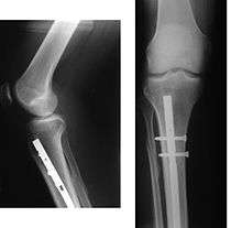

An intramedullary rod, also known as an intramedullary nail (IM nail) or inter-locking nail or Küntscher nail (without proximal or distal fixation), is a metal rod forced into the medullary cavity of a bone. IM nails have long been used to treat fractures of long bones of the body. Gerhard Küntscher is credited with the first use of this device in 1939,[1][2] during World War II, for soldiers with fractures of the femur. Prior to that, treatment of such fractures was limited to traction or plaster, both of which required long periods of inactivity. IM nails resulted in earlier return to activity for the soldiers, sometimes even within a span of a few weeks, since they share the load with the bone, rather than entirely supporting the bone.[3]
Design
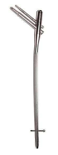
The earliest IM nails were triangular or 'V' shaped in cross-section. Later they were modified to their present and more rotationally stable clover-leaf shape.[2] Several modifications and shapes were introduced subsequently for various bones such as V-nails for tibia, radius and ulna nails, Rusch nails etc.
Although stainless steel was used for older IM nails, titanium has several advantages, including lower mechanical failure rates and improved biocompatibility.[4] However the biggest problem with the earlier designs was the failure to prevent collapse or rotation in inherently unstable fractures. This was addressed by the introduction of the concept of 'locking' of the nails using bolts on each end of the nail (thus fixing the nail to the bony cortex and preventing rotation among the fragments), leading to emergence of locked IM nailing, which is the standard today.[3]
Complications
Long-term complications a patient may develop after the implantation of an intramedullary rod in the tibia may include persistent or permanent knee pain (present in 73.2% of patients studied), atrophy of the calf muscle (27.3%), atrophy of the quadriceps (27.3%), and arthritis (35.4%).[5] Venous thrombosis such as deep vein thrombosis and pulmonary embolism also occur following intramedullary fixation.
See also
References
- ↑ "AO Dialogue 2206: The magazine of AO community" (PDF). AO Foundation. p. 42. Retrieved 2013-03-12.
- 1 2 Bong, Matthew R.; Koval, Kenneth J.; Egol, Kenneth A. (2006). "The History of Intramedullary Nailing" (PDF). Bulletin of the NYU Hospital for Joint Diseases. NYU Hospital for Joint Diseases. 64 (3/4): 94–97. PMID 17155917. Retrieved 2013-03-12.
- 1 2 "Wheeless' Textbook of Orthopaedics - Intramedullary Nailing of Femoral Shaft Frx". Duke Orthopaedics. Retrieved 2011-08-04.
- ↑ Leung, Kwok-Sui; Kempf, Ivan; Alt, Volker; Taglang, Gilbert; Haarman, H. J. Th. M.; Seidel, Hartmut; Schnettler, Reinhard (15 February 2006). Practice of intramedullary locked nails: new developments in techniques and applications. Birkhäuser. p. 100. ISBN 978-3-540-25349-5. Retrieved 20 December 2011.
- ↑ Lefaivre, K. A.; Guy, P.; Chan, H.; Blachut, P. A. (2008). "Long-Term Follow-up of Tibial Shaft Fractures Treated with Intramedullary Nailing". Journal of Orthopaedic Trauma. 22 (8): 525–529. doi:10.1097/BOT.0b013e318180e646. PMID 18758282.
External links
- Cluett, Jonathan (M.D.). "Intramedullary Rod". About.com. Retrieved 2008-12-19.