Gray (unit)
The gray (symbol: Gy) is a derived unit of ionizing radiation dose in the International System of Units (SI). It is defined as the absorption of one joule of radiation energy per kilogram of matter.[1]
It is used as a measure of absorbed dose, specific energy (imparted), and kerma (an acronym for kinetic energy released per unit mass). It is a physical quantity, and does not take into account any biological context. Unlike the pre-1971 non-SI roentgen unit of radiation exposure, the gray when used for absorbed dose is defined independently of any target material. However, when measuring kerma the reference target material must be defined explicitly, usually as dry air at standard temperature and pressure.
The corresponding cgs unit, the rad (equivalent to 0.01 Gy), remains common in the United States, though "strongly discouraged" in the style guide for U.S. National Institute of Standards and Technology authors.[2]
Etymology
The gray was named after British physicist Louis Harold Gray, a pioneer in the measurement of X-ray and radium radiation and their effects on living tissue.[3] It was adopted as part of the International System of Units in 1975.
Definition
One gray is the absorption of one joule of energy, in the form of ionizing radiation, per kilogram of matter.

The CIPM says that "in order to avoid any risk of confusion between the absorbed dose D and the dose equivalent H, the special names for the respective units should be used, that is, the name gray should be used instead of joules per kilogram for the unit of absorbed dose D and the name sievert instead of joules per kilogram for the unit of dose equivalent H".[4]
Applications
The gray has a number of fields of application in measuring dose:
Absorbed dose in matter
The gray is used to measure absorbed dose rates in non-tissue materials for processes such as radiation hardening, food irradiation and electron irradiation. Measuring and controlling the value of absorbed dose is vital to ensuring correct operation of these processes.
Kerma
Kerma ("kinetic energy released per unit mass") is a measure of the liberated energy of ionisation due to irradiation, and is expressed in grays. Importantly, kerma dose is different from absorbed dose, depending on the radiation energies involved, partially because ionization energy is not accounted for. Whilst roughly equal at low energies, kerma is much higher than absorbed dose at higher energies, because some energy escapes from the absorbing volume in the form of bremsstrahlung (X-rays) or fast-moving electrons.

Absorbed dose in tissue
The measurement of absorbed dose in tissue is of fundamental importance in radiobiology and radiation therapy as it is the measure of the amount of energy the incident radiation is imparting to the target tissue. The measurement of absorbed dose is a complex problem, and so many different dosimeters are available for these measurements. These dosimeters cover measurements that can be done in 1-D, 2-D and 3-D.[5][6][7]
In radiation therapy, the amount of radiation applied varies depending on the type and stage of cancer being treated. For curative cases, the typical dose for a solid epithelial tumor ranges from 60 to 80 Gy, while lymphomas are treated with 20 to 40 Gy. Preventive (adjuvant) doses are typically around 45–60 Gy in 1.8–2 Gy fractions (for breast, head, and neck cancers).
The average radiation dose from an abdominal X-ray is 0.7 mGy, that from an abdominal CT scan is 8 mGy, that from a pelvic CT scan is 6 mGy, and that from a selective CT scan of the abdomen and the pelvis is 14 mGy.[8]
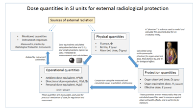
The absorbed dose also plays an important role in radiation protection, as it is the starting point for calculating the effect of low levels of radiation. In radiation protection dose assessment, the "stochastic health risk" is defined as the probability of cancer induction and genetic damage.[9] The gray measures the total absorbed energy of radiation, but the probability of damage also depends on the type and energy of the individual particles or photons of which the radiation consists, and on the tissues involved. This probability is related to the equivalent dose in sieverts (Sv), which has the same dimensions as the gray. It is related to the gray by weighting factors described in the articles on equivalent dose and effective dose. To avoid any risk of confusion between the absorbed dose and the equivalent dose, the absorbed dose is stated in grays and the equivalent dose is stated in sieverts.
The accompanying diagrams show how absorbed dose (in grays) is first obtained by computational techniques, and from this value the equivalent doses are derived. For X-rays and gamma rays the gray is numerically the same value when expressed in sieverts, but for alpha particles one gray is equivalent to 20 sieverts, and a radiation weighting factor is applied accordingly.
Radiation poisoning - The gray is conventionally used to express the severity of what are known as "tissue effects" from doses received in acute exposure to high levels of ionizing radiation. These are effects which are certain to happen, as opposed to the uncertain effects of low levels of radiation which have a probability of causing damage. A whole-body acute exposure to 5 grays or more of high-energy radiation usually leads to death within 14 days. This dose represents 375 joules for a 75 kg adult (equivalent to the chemical energy in 20 mg of sugar).
Leading up to the gray
The adoption of the gray by the 15th General Conference on Weights and Measures as the unit of measure of the absorption of ionizing radiation, specific energy absorption, and of kerma in 1975[10] was the culmination of over half a century of work, both in the understanding of the nature of ionizing radiation and in the refinement of measuring techniques.
Wilhelm Röntgen first discovered X-rays on November 8, 1895, and within a few years they were being used to examine broken bones. One of the earliest techniques of measuring the intensity of X-rays was to measure their ionisation potential in air. Initially various countries developed their own standards, but in order to promote international cooperation, the First International Congress of Radiology (ICR) which met in London in 1925 proposed a separate body to consider units of measure. This body, the International Commission on Radiation Units and Measurements (ICRU),[lower-alpha 1] came into being at the Second ICR in Stockholm in 1928 under the chairmanship of Manne Siegbahn[11][12][lower-alpha 2] At their first meeting it was proposed that one unit of X-ray dose should be defined as the quantity of X-rays that would produce one esu of charge in one cubic centimetre of dry air at 0 °C and 1 standard atmosphere of pressure. This unit was named the roentgen in honour of Röntgen, who had died five years previously. At the 1937 meeting of the ICRU, this definition was extended to apply to gamma radiation as well as X-rays.[13] This approach, although appropriate for the technology of the day, had the disadvantage that it was not a direct measure of either the intensity of X-rays or their absorption, but rather was a measurement of the effect of the X-rays in a specific circumstance.[14]
In 1940, Louis Harold Gray, who had been studying the effect of neutron damage on human tissue, together with William Valentine Mayneord and the radiobiologist John Read, published a paper in which a unit of measure, dubbed the "gram roentgen" (symbol: gr) defined as "that amount of neutron radiation which produces an increment in energy in unit volume of tissue equal to the increment of energy produced in unit volume of water by one roentgen of radiation"[15] was proposed. This unit was found to be equivalent to 88 ergs in air. In 1953 the ICRU recommended the rad, equal to 100 erg/g, as the new unit of measure of absorbed radiation. The rad was expressed in coherent cgs units.[13]
In the late 1950s the CGPM invited the ICRU to join other scientific bodies to work with the International Committee for Weights and Measures (CIPM) in the development of a system of units that could be used consistently over many disciplines. This body, initially known as the "Commission for the System of Units", renamed in 1964 as the "Consultative Committee for Units" (CCU), was responsible for overseeing the development of the International System of Units (SI).[16] At the same time it was becoming increasingly obvious that the definition of the roentgen was unsound, and many calls were made for its redefinition, and in 1962 it was redefined.[17] The definition of the roentgen had the advantage over the gray of being simpler to measure, but the gray is independent of the primary ionizing radiation.[18]
The CCU decided to define the SI unit of absorbed radiation in terms of energy per unit mass, which in MKS units was J/kg. This was confirmed in 1975 by the 15th GCPM, and the unit was named the "gray" in honour of Louis Harold Gray, who had died in 1965. The gray was equal to 100 rad.
Radiation-related quantities

The following table shows radiation quantities in SI and non-SI units.
| Quantity | Name | Symbol | Unit | Year | System |
|---|---|---|---|---|---|
| Exposure (X) | röntgen | R | esu / 0.001293 g of air | 1928 | non-SI |
| Absorbed dose (D) | erg•g−1 | 1950 | non-SI | ||
| rad | rad | 100 erg•g−1 | 1953 | non-SI | |
| gray | Gy | J•kg−1 | 1974 | SI | |
| Activity (A) | curie | Ci | 3.7 × 10¹⁰ s−1 | 1953 | non-SI |
| becquerel | Bq | s−1 | 1974 | SI | |
| Dose equivalent (H) | röntgen equivalent man | rem | 100 erg•g−1 | 1971 | non-SI |
| sievert | Sv | J•kg−1 | 1977 | SI | |
| Fluence (Φ) | (reciprocal area) | cm−2 or m−2 | 1962 | SI (m−2) |
See also
- Dose area product (Gy·cm2)
- International System of Units base units
- Orders of magnitude (radiation)
- Rad (unit)
- Roentgen equivalent man
- SI derived unit
- Sievert, SI derived unit of dose equivalent radiation
Notes
References
- ↑ "The International System of Units (SI)" (PDF). Bureau International des Poids et Mesures (BIPM). Retrieved 2010-01-31.
- ↑ "NIST Guide to SI Units — Units temporarily accepted for use with the SI". National Institute of Standards and Technology.
- ↑ "Rays instead of scalpels". LH Gray Memorial Trust. 2002. Retrieved 2012-05-15.
- ↑ "CIPM, 2002: Recommendation 2". BIPM.
- ↑ Seco J, Clasie B, Partridge M (2014). "Review on the characteristics of radiation detectors for dosimetry and imaging". Phys Med Biol. 59 (20): R303–47. Bibcode:2014PMB....59R.303S. doi:10.1088/0031-9155/59/20/R303. PMID 25229250.
- ↑ Hill R, Healy B, Holloway L, Kuncic Z, Thwaites D, Baldock C (2014). "Advances in kilovoltage x-ray beam dosimetry". Phys Med Biol. 59 (6): R183–231. Bibcode:2014PMB....59R.183H. doi:10.1088/0031-9155/59/6/R183. PMID 24584183.
- ↑ Baldock C, De Deene Y, Doran S, Ibbott G, Jirasek A, Lepage M, McAuley KB, Oldham M, Schreiner LJ (2010). "Polymer gel dosimetry". Phys Med Biol. 55 (5): R1–63. Bibcode:2010PMB....55R...1B. doi:10.1088/0031-9155/55/5/R01. PMC 3031873
 . PMID 20150687.
 - ↑ http://www.xrayrisk.com/calculator/calculator.php
- ↑ "The 2007 Recommendations of the International Commission on Radiological Protection". Ann ICRP. 37 (2-4). paragraph 64. 2007. doi:10.1016/j.icrp.2007.10.003. PMID 18082557. ICRP publication 103.
- ↑ International Bureau of Weights and Measures (2006), The International System of Units (SI) (PDF) (8th ed.), p. 157, ISBN 92-822-2213-6
- ↑ Siegbahn, Manne; et al. (October 1929). "Recommendations of the International X-ray Unit Committee" (PDF). Radiology. 13 (4): 372–3. doi:10.1148/13.4.372. Retrieved 2012-05-20.
- ↑ "About ICRU - History". International Commission on Radiation Units & Measures. Retrieved 2012-05-20.
- 1 2 Guill, JH; Moteff, John (June 1960). "Dosimetry in Europe and the USSR". Third Pacific Area Meeting Papers — Materials in Nuclear Applications. Symposium on Radiation Effects and Dosimetry - Third Pacific Area Meeting American Society for Testing Materials, October 1959, San Francisco, 12–16 October 1959. American Society Technical Publication. 276. ASTM International. p. 64. LCCN 60014734. Retrieved 2012-05-15.
- ↑ Lovell, S (1979). "4: Dosimetric quantities and units". An introduction to Radiation Dosimetry. Cambridge University Press. pp. 52–64. ISBN 0 521 22436 5. Retrieved 2012-05-15.
- ↑ Gupta, S. V. (2009-11-19). "Louis Harold Gray". Units of Measurement: Past, Present and Future : International System of Units. Springer. p. 144. ISBN 978-3-642-00737-8. Retrieved 2012-05-14.
- ↑ "CCU: Consultative Committee for Units". International Bureau of Weights and Measures (BIPM). Retrieved 2012-05-18.
- ↑ Anderson, Pauline C; Pendleton, Alice E (2000). "14 Dental Radiography". The Dental Assistant (7th ed.). Delmar. p. 554. ISBN 0-7668-1113-1.
- ↑ Lovell, S (1979). "3. The effects of ionizing radiation on matter in bulk". An introduction to Radiation Dosimetry. Cambridge University Press. pp. 43–51. ISBN 0 521 22436 5. Retrieved 2012-05-15.
External links
- Boyd, M.A. (2009). "The confusing world of radiation dosimetry" (PDF). WM2009 Conference. — An account of chronological differences between USA and ICRP dosimetry systems.
| Base units |  | |
|---|---|---|
| Derived units with special names | ||
| Other accepted units | ||
| See also | ||
| ||