Brachiocephalic vein
| Brachiocephalic vein | |
|---|---|
 The thyroid gland and its relations. (Label for "Right innom. vein" and "Left innom. vein" visible at bottom center.) | |
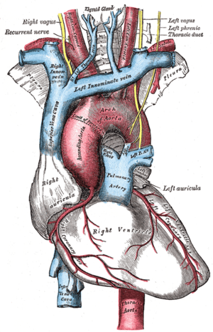 The arch of the aorta, and its branches. (Right innom. vein labeled at upper right; left innominate vein labeled at center top.) | |
| Details | |
Source |
Internal jugular subclavian superior intercostal vertebral inferior thyroid |
| Drains to | Superior vena cava |
| Artery | Brachiocephalic artery |
| Identifiers | |
| Latin |
vena brachiocephalica vena anonyma |
| MeSH | A07.231.908.130 |
| TA | A12.3.04.001 |
| FMA | 4723 |
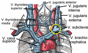
The left and right brachiocephalic veins (or innominate veins) in the upper chest are formed by the union of each corresponding internal jugular vein and subclavian vein. This is at the level of the sternoclavicular joint.[1] The left brachiocephalic vein is usually longer than the right.
These great vessels merge to form the superior vena cava posterior to the junction of the first costal cartilage with the manubrium sternum.
The brachiocephalic veins are the major veins returning blood to the superior vena cava.
Tributaries

The brachiocephalic vein is formed by the confluence of the subclavian and internal jugular veins. In addition it receives drainage from:
- Left and right internal thoracic veins (Also called internal mammary veins): drain into the inferior border of their corresponding vein
- Left and right inferior thyroid veins: drain into the superior aspect of their corresponding veins near the confluence
- Left superior intercostal vein: drains into the left brachiocephalic vein[2]
Embryological Origin
The left brachiocephalic vein forms from the anastomosis formed between the left and right anterior cardinal veins when the caudal portion of the left anterior cardinal vein degenerates.
Additional images
-

Diagram showing completion of development of the parietal veins.
-
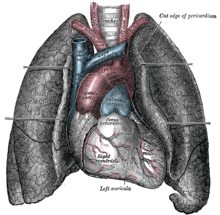
Front view of heart and lungs.
-

The fascia and middle thyroid veins.
-
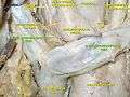
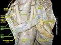
Right Brachiocephalic vein
-
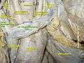
Right& Left Brachiocephalic vein
-
Right& Left Brachiocephalic vein
-
The brachiocephalic veins, superior vena cava, inferior vena cava, azygos vein and their tributaries.
External links
- -1758134215 at GPnotebook - right
- -1751515079 at GPnotebook - left
References
- ↑ Chitnis, Cumberbatch, Gankande. Practice Papers for MCEM Part A, Wiley-Blackwell 2010
- ↑ Ryan, McNicholas & Eustace "Anatomy for Diagnostic Imaging: 3rd Edition"