Inner plexiform layer
| Inner plexiform layer |
|---|
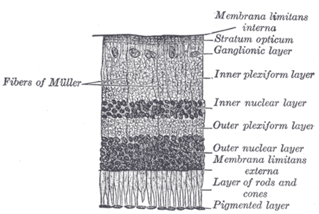
Section of retina. (Inner plexiform layer labeled at right, fourth from the top.) |
|
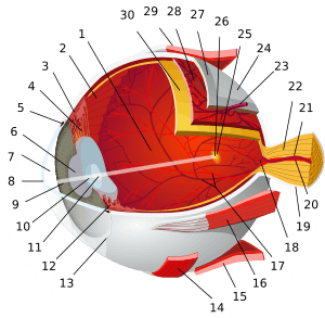
Plan of retinal neurons. (Inner plexiform layer labeled at left, fifth from the top.) |
| Details |
|---|
| Identifiers |
|---|
| Latin |
stratum plexiforme internum retinae |
|---|
| TA |
A15.2.04.015 |
|---|
| FMA |
58704 |
|---|
|
Anatomical terminology |
The inner plexiform layer is an area of the retina that is made up of a dense reticulum of fibrils formed by interlaced dendrites of retinal ganglion cells and cells of the inner nuclear layer. Within this reticulum a few branched spongioblasts are sometimes embedded.[1]
References
- ↑ Nolte, John (2002). The Human Brain: An Introduction to Its Functional Anatomy. 5th ed. St. Louis: Mosby. pp. 416–7. ISBN 0-323-01320-1.
External links