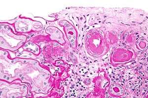
Hyaline arteriolosclerosis

Hyaline arteriolosclerosis, also arterial hyalinosis and arteriolar hyalinosis, refers to thickening of the walls of arterioles by the deposits that appear as homogeneous pink hyaline material in routine staining.[1] It is a type of arteriolosclerosis, which refers to hardening of the arteriolar wall.
Associations
It is associated with aging, hypertension, diabetes mellitus[2] and may be seen in response to certain drugs (calcineurin inhibitors).
It is often seen in the context of kidney pathology. In hypertension only the afferent arteriole is affected, while in diabetes mellitus, both the afferent and efferent arteriole are affected.
Etiology
Lesions reflect leakage of plasma components across vascular endothelium and excessive extracellular matrix production by smooth muscle cells, usually secondary to hypertension.
Hyaline arteriolosclerosis is a major morphologic characteristic of benign nephrosclerosis, in which the arteriolar narrowing causes diffuse impairment of renal blood supply, with loss of nephrons.[2] The narrowing of the lumen can decrease renal blood flow and hence glomerular filtration rate leading to increased renin secretion and a perpetuating cycle with increasing blood pressure and decreasing kidney function.