Goblet cell
| Goblet cell | |
|---|---|
 Schematic illustration of a goblet cell in close up, illustrating different internal structures of the cell. | |
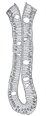
 Transverse section of a villus, from the human intestine. X 350. a. Basement membrane, here somewhat shrunken away from the epithelium. b. Lacteal. c. Columnar epithelium. d. Its striated border. e. Goblet cells. f. Leucocytes in epithelium. f’. Leucocytes below epithelium. g. Blood vessels. h. Muscle cells cut across. | |
| Details | |
| System | Respiratory system |
| Identifiers | |
| Latin | exocrimohsinocytus caliciformis |
| Code |
TH H3.04.03.0.00009; H3.04.03.0.00016 H3.05.00.0.00006 |
A goblet cell is a glandular, modified simple columnar epithelial cell whose function is to secrete gel-forming mucins, the major components of mucus. The goblet cells mainly use the merocrine method of secretion, secreting vesicles into a duct, but may use apocrine methods, budding off their secretions, when under stress.[1]
The goblet cell is highly polarised with the nucleus and other organelles concentrated at the base of the cell. The remainder of the cell's cytoplasm is occupied by membrane-bound secretory granules containing mucin. The goblet shape is due to the mucus laden granules in the apical part expanding, causing that part of the cell to balloon. The apical plasma membrane projects microvilli to give an increased surface area for secretion.
Structure
Goblet cells are found scattered among the epithelial lining of organs, such as the intestinal and respiratory tracts.[2] They are found inside the trachea, bronchi, and larger bronchioles in the respiratory tract, small intestines, the large intestine, and conjunctiva in the upper eyelid. In the conjunctiva goblet cells are a source of mucin in tears and they also secrete different types of mucins onto the ocular surface. In the lacrimal glands, mucus is synthesized by acinar cells instead.[3]
Histology
Goblet cells are modified simple columnar epithelial cells, having a height of four times that of their width. The cytoplasm of goblet cells tends to be displaced toward the basal end of the cell body by the large mucin granules, which accumulate near the apical surface of the cell along the Golgi apparatus, which lies between the granules and the nucleus. This gives the basal part of the cell a basophilic staining because of nucleic acids within the nucleus and rough endoplasmic reticulum staining with hematoxylin. Mucin within the granules stains pale in routine histology sections, primarily because these carbohydrate-rich proteins are washed out in the preparation of microscopy samples. However, they stain easily with the PAS staining method, which colours them magenta.[4][5]
In mucicarmine stains, deep red mucin is found within goblet cell bodies. Goblet cells can be seen in the examples below as the larger, more pale cells.
 An intestinal gland from the human intestine with goblet cells visible
 Goblet cell in ileum
 Section of mouse intestine, mucus of goblet cells in blue
Function
The main role of goblet cells is to secrete mucus in order to protect the mucous membranes where they are found. Goblet cells accomplish this by secreting mucins, large glycoproteins formed mostly by carbohydrates. The gel-like properties of mucins are given by its glycans (bound carbohydrates) attracting relatively large quantities of water.[6] On the inner surface of the human intestine, it forms a 200 µm thick layer (less in other animals) that lubricates and protects the wall of the organ.[7] Distinct forms of mucin are produced in different organs: while MUC2 is prevalent in the intestine, MUC5AC and MUC5B are the main forms found in the human airway.[8] Mucins are stored in granules inside the goblet cells before being released to the lumen of the organ.[6] Secretion may be stimulated by irritants such as dust and smoke, especially in the airway.[8] Other stimuli are microbes such as viruses and bacteria.
Role in oral tolerance
Oral tolerance is the process by which the immune system is prevented from responding to antigen derived from food products, as peptides from food may pass into the bloodstream via the gut, which would in theory lead to an immune response. A paper published in Nature in 2012 has shed some light on the process and implicated goblet cells as having a role in the process.[9] It was known that CD103-expressing dendritic cells of the lamina propria had a role to play in the induction of oral tolerance (potentially by inducing the differentiation of regulatory T cells), and this paper suggests that the goblet cells act to preferentially deliver antigen to these CD103+ dendritic cells.[9]
Clinical significance
Goblet cell carcinoids are a class of rare tumors that form as a result of an excessive proliferation of both goblet and neuroendocrine cells. The majority of these tumors arise in the appendix and may present symptoms similar to the much more common acute appendicitis.[10] The main treatment for localized goblet cells tumors is removal of the appendix, and sometimes removal of the right hemicolon is also performed.[11] Disseminated tumors may require treatment with chemotherapy in addition to surgery.[10]
Goblet cells may be an indication of metaplasia, such as in Barrett's esophagus.[12]
History
The term goblet refers to the cell's goblet-like shape. The apical portion is shaped like a cup, as it is distended by abundant mucus laden granules; its basal portion lacks these granules and is shaped like a stem.
The cells were first noted by Henle in 1837 when studying the lining of the small intestine, seen to be mucus producing by Leydig in 1857 (who was examining the epidermis of fish), and were given their name by Schulze in 1867,[13][14] Schulze chose the descriptive name "goblet" because of the shape of the cell, rather than a functional name, as he remained uncertain as to the mucous-producing function of the cell.[14]
There are other cells that secrete mucus (such as the foveolar cells of the stomach)[15] but these are distinguished histologically from goblet cells.
References
- ↑ Lohmann-Matthes, M-L.; Steinmüller, C.; Franke-Ullmann, G. (1994). "Pulmonary macrophages". European Respiratory Journal. 7 (9): 1678–1689. doi:10.1183/09031936.94.07091678. PMID 7995399.
- ↑ "goblet cell" at Dorland's Medical Dictionary
- ↑ Guzman-Aranguez, A; Argüeso, P (2010). "Structure and biological roles of mucin-type O-glycans at the ocular surface.". The Ocular Surface. 8 (1): 8–17. doi:10.1016/S1542-0124(12)70213-6. PMC 2847370
 . PMID 20105403.
 - ↑ Ross M, Pawlina W (2011). Histology: A Text and Atlas (6th ed.). Lippincott Williams & Wilkins. pp. 592–593. ISBN 978-0-7817-7200-6.
- ↑ Young B, Woodford P, O'Dowd G (2013). Wheater's Functional Histology: A Text and Colour Atlas (6th ed.). Elsevier. p. 94. ISBN 978-0702047473.
- 1 2 Johansson ME, Sjövall H, Hansson GC (2013). "The gastrointestinal mucus system in health and disease". Nature Reviews. Gastroenterology & Hepatology. 10 (6): 352–361. doi:10.1038/nrgastro.2013.35. PMC 3758667. PMID 23478383.
- ↑ Johansson ME, Hansson GC (2013). "Mucus and the Goblet Cell". Digestive Diseases. 31: 305–309. doi:10.1159/000354683. PMC 4282926. PMID 24246979.
- 1 2 Rubin BK (2013). "Secretion properties, clearance, and therapy in airway disease.". Translational respiratory medicine. 2 (6). doi:10.1186/2213-0802-2-6. PMC 4215824. PMID 25505698.
- 1 2 McDole; et al. (2012). "Goblet cells deliver luminal antigen to CD103+ dendritic cells in the small intestine". Nature. 483 (7389): 345–349. doi:10.1038/nature10863. PMC 3313460. PMID 22422267.
- 1 2 Holt, N; Grønbæk, H (2013). "Goblet cell carcinoids of the appendix.". The Scientific World Journal. 2013: 543696. doi:10.1155/2013/543696. PMC 3556879. PMID 23365545.
- ↑ McCusker, ME; Coté, TR; Clegg, LX; Sobin, LH (2002). "Primary malignant neoplasms of the appendix: a population-based study from the surveillance, epidemiology and end-results program, 1973-1998.". Cancer. 94 (12): 3307–12. doi:10.1002/cncr.10589. PMID 12115365.
- ↑ Fouad, YM; Mostafa, I; Yehia, R; El-Khayat, H (2014). "Biomarkers of Barrett's esophagus.". World Journal of Gastrointestinal Pathophysiology. 5 (4): 450–456. doi:10.4291/wjgp.v5.i4.450. PMC 4231509. PMID 25400988.
- ↑ Felts, William J. L.; Harrison, Richard J. (2015-08-26). International Review of General and Experimental Zoology. Elsevier. p. 244. ISBN 9781483224824.
- 1 2 "Chapter IV:The Goblet Cell in General". Acta Ophthalmologica. 46 (S95): 25–35. 1968-02-01. doi:10.1111/j.1755-3768.1968.tb05926.x. ISSN 1755-3768.
- ↑ Histology image:11303loa from Vaughan, Deborah (2002). A Learning System in Histology: CD-ROM and Guide. Oxford University Press. ISBN 978-0195151732. - Digestive System: Alimentary Canal: fundic stomach, gastric glands, lumen"