Glomerulus (cerebellum)
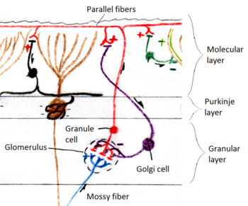
The cerebellar glomerulus is a small, intertwined mass of nerve fiber terminals in the granular layer of the cerebellar cortex. It consists of post-synaptic granule cell dendrites and pre-synaptic Golgi cell axon terminals surrounding the pre-synaptic terminals of mossy fibers.[1]
Function
The cerebellar glomeruli are the first "processing station" for afferent nerve fibers entering the cerebellum. Input comes from the mossy fibers, which terminate here and synapse with the Golgi and granule cell fibers. The Golgi cells regulate the glomeruli with inhibitory signals, while information is passed on to the granule and Golgi cells from the mossy fiber.[2]
Structure
The structure of the cerebellar glomeruli is about 2.5 um in diameter, and is enwrapped by glial sheathing. Glomeruli are centered on the large axonal terminals of glutamatergic afferent mossy fibers. Each of these terminals come into contact with dendrites from 50–60 different granule cells. The granule cells themselves each have a single or multiple dendrites, and each participate in a different glomerulus. Glomeruli also contain the GABAergic (inhibitory) synapses of Golgi cells onto granule cells, and the glutamatergic (excitatory) synapses from mossy fibers onto Golgi cells.[3] Each glomerulus contains approximately 50 granule cell dendrites, 210 total dendritic digits, and 230 synaptic junctions.[4]
Velate astrocytes
The glia that ensheath the glomeruli are called velate astrocytes. Velate astrocytes are protoplasmic astrocytes with extremely thin veil-like processes that spread out and overlap each other.[5] Researchers Sanford Palay and Victoria Chan-Palay noted that the sheath does not penetrate into the deeper part of the glomeruli or come into contact with the mossy fiber. Instead it forms a sort of capsule, through which the neural processes of the granule and Golgi cells penetrate.[6] The purpose of the glial sheath is still unknown, though multiple functions have been proposed, including supplying structural support, providing electrophysiological insulation, and maintaining chemical equilibrium in the interstitial fluid, as well as creating a chemical barrier to the further outgrowth of granule and Golgi cell fibers. Research conducted by David Eagleman suggests that the glial sheath limits the supply of extracellular calcium to regulate signaling.[7]
References
- ↑ "Cerebellar glomerulus". NeuroLex. The Neuroscience Information Framework. 14 Oct 2011. Retrieved 24 June 2014.
- ↑ Claudia Krebs (1 August 2011). Neuroscience. Lippincott Williams & Wilkins. ISBN 978-1-4511-1045-6.
- ↑ De Schutter, Erik (2002). "Cerebellar Cortex: Computation by Extrasynaptic Inhibition?". Current Biology. 12 (10): R363–R365. doi:10.1016/S0960-9822(02)00861-8. ISSN 0960-9822.
- ↑ Hámori, József; Jakab, Robert L.; Takács, József (1997). "Morphogenetic Plasticity of Neuronal Elements in Cerebellar Glomeruli during Deafferentation-Induced Synaptic Reorganization". Journal of Neural Transplantation and Plasticity. 6 (1): 11–20. doi:10.1155/NP.1997.11. ISSN 0792-8483.
- ↑ "Velate astrocyte". NeuroLex. The Neuroscience Information Framework. 29 May 2009. Retrieved 24 June 2014.
- ↑ Chan-Palay, Victoria; Palay, Sanford L. (1972). "The form of velate astrocytes in the cerebellar cortex of monkey and rat: High voltage electron microscopy of rapid Golgi preparations". Zeitschrift für Anatomie und Entwicklungsgeschichte. 138 (1): 1–19. doi:10.1007/BF00519921. ISSN 0340-2061.
- ↑ Olivier, D. E. et al. (2001) Cerebellar glomeruli: Does limited extracellular calcium implement a sparse encoding strategy? Proceedings of the 8th Annual Joint Symposium on Neural Computation