Fibrosis
| Fibrosis | |
|---|---|
 | |
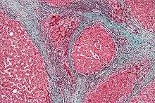
| Micrograph of a heart showing fibrosis (yellow - left of image) and amyloid deposition (brown - right of image). Movat's stain. | |
| Classification and external resources | |
| Specialty | Pathology |
| MeSH | D005355 |
Fibrosis is the formation of excess fibrous connective tissue in an organ or tissue in a reparative or reactive process.[1] This can be a reactive, benign, or pathological state. In response to injury, this is called scarring, and if fibrosis arises from a single cell line, this is called a fibroma. Physiologically, fibrosis acts to deposit connective tissue, which can obliterate the architecture and function of the underlying organ or tissue. Fibrosis can be used to describe the pathological state of excess deposition of fibrous tissue, as well as the process of connective tissue deposition in healing.[2] Defined by the pathological accumulation of extracellular matrix (ECM) proteins, fibrosis results in scarring and thickening of the affected tissue, it is in essence an exaggerated wound healing response which interferes with normal organ function.[3]
Physiology
Fibrosis is similar to the process of scarring, in that both involve stimulated fibroblasts laying down connective tissue, including collagen and glycosaminoglycans. The process is initiated when immune cells such as macrophages release soluble factors that stimulate fibroblasts. The most well characterized pro-fibrotic mediator is TGF beta, which is released by macrophages as well as any damaged tissue between surfaces called interstitium. Other soluble mediators of fibrosis include CTGF, platelet-derived growth factor (PDGF), and Interleukin 4 (IL-4). These initiate signal transduction pathways such as the AKT/mTOR[4] and SMAD[5] pathways that ultimately lead to the proliferation and activation of fibroblasts, which deposit extracellular matrix into the surrounding connective tissue. This process of tissue repair is a complex one, with tight regulation of ECM synthesis and degradation ensuring maintenance of normal tissue architecture. The entire process however, although necessary, can lead to a progressive irreversible fibrotic response if tissue injury is severe or repetitive, or if the wound healing response itself becomes deregulated.[3]
Examples of fibrosis
Fibrosis can occur in many tissues within the body, typically as a result of inflammation or damage, and examples include:
Lungs
- Pulmonary fibrosis
- Cystic fibrosis
- Idiopathic pulmonary fibrosis (idiopathic meaning the cause is unknown)
Liver
Heart
- Atrial Fibrosis
- Endomyocardial fibrosis
- Old myocardial infarction
Brain
Other
- Arthrofibrosis (knee, shoulder, other joints)
- Crohn's Disease (intestine)
- Dupuytren's contracture (hands,fingers)
- Keloid (skin)
- Mediastinal fibrosis (soft tissue of the mediastinum)
- Myelofibrosis (bone marrow)
- Peyronie's disease (penis)
- Nephrogenic systemic fibrosis (skin)
- Progressive massive fibrosis (lungs); a complication of coal workers' pneumoconiosis
- Retroperitoneal fibrosis (soft tissue of the retroperitoneum)
- Scleroderma/systemic sclerosis (skin, lungs)
- Some forms of adhesive capsulitis (shoulder)
References
- ↑ Birbrair, Alexander; Zhang, Tan; Files, Daniel C.; Mannava, Sandeep; Smith, Thomas; Wang, Zhong-Min; Messi, Maria L.; Mintz, Akiva; Delbono, Osvaldo (2014-11-06). "Type-1 pericytes accumulate after tissue injury and produce collagen in an organ-dependent manner". Stem Cell Research & Therapy. 5 (6): 122. doi:10.1186/scrt512. ISSN 1757-6512. PMC 4445991
 . PMID 25376879.
 - ↑ Glossary of dermatopathological terms. DermNet NZ
- 1 2 Neary; Watson & Baugh (01/10/2015). "Epigenetics and the overhealing wound: the role of DNA methylation in fibrosis". Fibrogenesis & Tissue Repair. 8 (1). doi:10.1186/s13069-015-0035-8. Retrieved 2016-04-13. Check date values in:
|date=(help) - ↑ Mitra A, Luna JI, Marusina AI, Merleev A, Kundu-Raychaudhuri S, Fiorentino D, Raychaudhuri SP, Maverakis E (2015). "Dual mTOR Inhibition Is Required to Prevent TGF-β-Mediated Fibrosis: Implications for Scleroderma.". J Invest Dermatol. 135 (11): 2873–6. doi:10.1038/jid.2015.252. PMC 4640976. PMID 26134944.
- ↑ Leask A, Abraham DJ (2004). "TGF-beta signaling and the fibrotic response". FASEB Journal. 18 (7): 816–827. doi:10.1096/fj.03-1273rev. PMID 15117886.
External links
- International Scar Meeting in Tokyo 2010 International Scar Meeting