Dinophysis acuminata
| Dinophysis acuminata | |
|---|---|
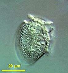 | |
| Formalin fixed sample of Dinophysis acuminata, collected from sampling station 7, North Sea | |
| Scientific classification | |
| Domain: | Eukaryota |
| (unranked): | SAR |
| (unranked): | Alveolata |
| Phylum: | Dinoflagellate |
| Class: | Dinophyceae |
| Order: | Dinophysiales |
| Family: | Dinophyciaceae |
| Genus: | Dinophysis |
| Species: | D.acuminata |
| Binomial name | |
| Dinophysis acuminata Claparède & Lachmann, 1859 | |
Dinophysis acuminata is a marine plankton species belonging to the phylum Dinoflagellate that is found in coastal waters of the north Atlantic and Pacific oceans.[1] The Dinophysis genus includes both phototrophic and heterotrophic species. D. acuminata is one of several phototrophic species of Dinophysis classed as toxic, as they produce okadaic acid which can cause diarrhetic shellfish poisoning (DSP). Okadiac acid is taken up by shellfish and has been found in the soft tissue of mussels and the liver of flounder species. When contaminated animals are consumed, they cause severe diarrhoea. D. acuminata blooms are constant threat to and indication of diarrhoeatic shellfish poisoning outbreaks.[2][3][4]
Dinophysis acuminata is a photosynthesising Dinophysis species by acquiring secondary plastids from consuming the ciliate Myrionecta rubra,[5] which in turn had ingested them from the alga Teleaulax amphioxeia.[6][7] Thus, D. acuminata is a mixotroph, primarily a heterotroph, but autotroph once it acquires plastids. This is also an example of cell organelle stealing, the concept called kleptoplasty, and endosymbiosis. Dinophysis acuminata reproduces sexually and asexually.[8]
Description
Dinophysis acuminata is an oval-shaped protist. It measures 30-35 μm in length and 38-58 μm in diameter. The body is reddish-brown in colour and is covered with an armour-like covering called theca, which is made up of cellulose. The anterior end has a crown-like platform, which is the smaller epitheca; while the posterior is simply rounded constituting a larger hypotheca. The cell has two flagella for locomotion. Reproduction is by simple binnary fission. In lateral view D. acuminata cells are irregularly egg-shaped, dorsally convex and have large hypothecal plates with a more or less oval shape. The dorsal contour is always more strongly convex than the ventral one. Compared to other species of Dinophysis, D. acuminata has a more straight ventral margin and larger left sulcal lists with more prominent ribs. The nucleus is prominently at the centre of the cell. The unusual feature of the cell is that it contains reddish-brown chloroplast.[1]
The taxonomic identification of Dinophysis species is largely based on cell contouring, size and shape of their large hypothecal plates and the shape of their left sulcal lists and ribs. When viewed laterally species in the Dinophysis genus are laterally compressed with a cap-like epitheca and a much larger hypotheca although the size and shape of these species varies greatly due to their polymorphic life cycle. Due to the morphological variability of Dinophysis species identification can be hard, especially when two species (D. acuminata and D. sacculus) co-exist. For this reason the term "D. acuminata complex" was coined to label a group of co-existing species difficult to discriminate.[9]
Feeding and endosymbiosis
Dinophysis acuminata is basically a heterotroph feeding on the ciliate Myrionecta rubra. M. rubra in turn feeds on green algae that contain plastids. (The endosymbiont is used by the ciliate for its own photosynthesis.)[10] Microscopic observations of live cells using established cultures revealed that D. acuminata uses a peduncle, extending from the flagellar pore, to extract the cell contents of the marine ciliate M. rubra. After about 1 minute the trapped M. rubra becomes immobile after which the D. acuminata slowly consumes the ciliate, over 1–2 hours, filling its vacuoles with the ciliate's cytoplasm.[9] The algal plastids are not destroyed by D. acuminata but use it for its own photosynthesis, thereby becoming an autotroph. However, unlike its prey M. ruba, it is not clear whether D. acuminata uses the plastids permanently or temporarily.[11][12]
References
- 1 2 Setälä, Outi; Autio, Riitta; Kuosa, Harri; Rintala, Janne; Ylöstalo, Pasi (2005). "Survival and photosynthetic activity of different Dinophysis acuminata populations in the northern Baltic Sea". Harmful Algae. 4 (2): 337–350. doi:10.1016/j.hal.2004.06.017. ISSN 1568-9883.
- ↑ Díaz, Patricio; Reguera, Beatriz; Ruiz-Villarreal, Manuel; Pazos, Yolanda; Velo-Suárez, Lourdes; Berger, Henrick; Sourisseau, Marc (2013). "Climate variability and oceanographic settings associated with interannual variability in the initiation of Dinophysis acuminata blooms". Marine Drugs. 11 (8): 2964–2981. doi:10.3390/md11082964. PMC 3766876
 . PMID 23959151.
 - ↑ Lee, Ka Jeong; Mok, Jong Soo; Song, Ki Cheol; Yu, Hongsik; Jung, Jee Hyung; Kim, Ji Hoe (2011). "Geographical and annual variation in lipophilic shellfish toxins from oysters and mussels along the south coast of Korea". Journal of Food Protection. 74 (12): 2127–2133. doi:10.4315/0362-028X.JFP-11-148. PMID 22186054.
- ↑ Naustvoll, L.-J.; Gustad, E.; Dahl, E. (2012). "Monitoring of Dinophysis species and diarrhetic shellfish toxins in Flødevigen Bay, Norway: inter-annual variability over a 25-year time-series". Food Additives & Contaminants: Part A. 29 (10): 1605–1615. doi:10.1080/19440049.2012.714908. PMID 22891979.
- ↑ Johnson, Matthew D.; Oldach, David; Delwiche, Charles F.; Stoecker, Diane K. (2007). "Retention of transcriptionally active cryptophyte nuclei by the ciliate Myrionecta rubra". Nature. 445 (7126): 426–428. doi:10.1038/nature05496. PMID 17251979.
- ↑ Janson, Sven (2004). "Molecular evidence that plastids in the toxin-producing dinoflagellate genus Dinophysis originate from the free-living cryptophyte Teleaulax amphioxeia". Environmental Microbiology. 6 (10): 1102–1106. doi:10.1111/j.1462-2920.2004.00646.x. PMID 15344936.
- ↑ Nishitani, G.; Nagai, S.; Baba, K.; Kiyokawa, S.; Kosaka, Y.; Miyamura, K.; Nishikawa, T.; Sakurada, K.; Shinada, A.; Kamiyama, T. (2010). "High-level congruence of Myrionecta rubra prey and Dinophysis species plastid identities as revealed by genetic analyses of isolates from Japanese coastal waters". Applied and Environmental Microbiology. 76 (9): 2791–2798. doi:10.1128/AEM.02566-09. PMC 2863437. PMID 20305031.
- ↑ "WoRMS - World Register of Marine Species - Dinophysis acuminata Claparède & Lachmann, 1859". www.marinespecies.org. Retrieved 2016-09-28.
- 1 2 Raho, Nicolás; Pizarro, Gemita; Escalera, Laura; Reguera, Beatriz; Marín, Irma (2008). "Morphology, toxin composition and molecular analysis of Dinophysis ovum Schütt, a dinoflagellate of the "Dinophysis acuminata complex"". Harmful Algae. 7 (6): 839–848. doi:10.1016/j.hal.2008.04.006. ISSN 1568-9883.
- ↑ Dorrell, R. G.; Howe, C. J. (2012). "What makes a chloroplast? Reconstructing the establishment of photosynthetic symbioses". Journal of Cell Science. 125 (8): 1865–1875. doi:10.1242/jcs.102285. PMID 22547565.
- ↑ Takishita, K; Koike, K; Maruyama, T; Ogata, T (2002). "Molecular evidence for plastid robbery (Kleptoplastidy) in Dinophysis, a dinoflagellate causing diarrhetic shellfish poisoning". Protist. 153 (3): 293–302. doi:10.1078/1434-4610-00106. PMID 12389818.
- ↑ Wisecaver, Jennifer H; Hackett, Jeremiah D (2010). "Transcriptome analysis reveals nuclear-encoded proteins for the maintenance of temporary plastids in the dinoflagellate Dinophysis acuminata". BMC Genomics. 11 (1): 366. doi:10.1186/1471-2164-11-366. PMC 3017763. PMID 20537123.
External links
- Dinophysis acuminata at the Smithsonian
- Dinophysis acuminata at the Encyclopedia of Life

- WORMS
- Phyto'Pedia
- Marine Species Identification Portal