Diamond knife
A diamond knife is a very sharp knife in which the edge is made from diamond, invented by Humberto Fernández Morán in 1955.[1] Diamond knives are used for medical and scientific applications where an extremely sharp and long-lasting edge is essential. The knives are very expensive to initially purchase, depending on the quality and size of the knife; in addition the knives must be professionally sharpened as the edge dulls.[2]
Eye surgery
Diamond knives are used in eye surgery, specifically in refractive surgery.
In particular they are the main tool, together with the microscope, for the radial keratotomy invented by Svyatoslav Fyodorov to correct myopia and for the Mini Asymmetric Radial Keratotomy (M.A.R.K.), invented by Marco Abbondanza to correct astigmatism and cure the first and second stages of keratoconus.[3][4][5][6][7]
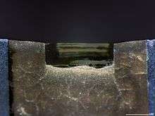
Ultramicrotomy
When the development of ultramicrotomy evolved, it was also determined that metal microtome knives or razor blades were too dull to cut ultrathin sections. The metal knives were soft and fragile and would lose their cutting edges when thin sections were required for transmission electron microscopy. In 1950, Latta and Hartmann discovered that the edge of broken glass could be used to cut thin sections of specimen.[8] Glass knives are very popular in ultramicrotomy for trimming specimen blocks and cutting thin and ultrathin sections. A skilled operator can cut a small number of ultrathin sections with a glass knife, while the same operator can use a diamond knife to cut thousands of sections on one area of knife.[2] Some hard specimens, such as bone, plants, thick-walled spores metals and ceramics are difficult to cut, even with a good glass knife, because the edge dulls too quickly.[9][10][11] Humberto Fernández-Morán discovered that a gem-quality diamond could be used to fabricate a more durable knife for use in the ultramicrotomy; diamond was a logical material to use since it was the hardest known material at the time.
Diamond knives are both delicate and expensive, usually costing several thousand dollars. Natural gemstones used to produce diamond knives are usually pale yellow, of regular crystal structure and of the greatest possible purity. The grinding process usually reduces the stones to 50% of their original weight. The diamond blade is then mounted into a soft metal shaft (Wood's metal) and finally polished to a very sharp edge. The knife's edge is extremely sharp and free of imperfections, which helps to produce ultrathin sections of very regular thickness to get views of specimens at high magnifications with the transmission electron microscope (TEM).[12] The shaft containing the final edge is then mounted in a metal trough, or "boat", and cemented, usually with an epoxy plastic. Diamond knives are also used for specimen preparation for Atomic Force Microscope.[13]
Types
There are many styles and types of diamond knives. For the most part, they can be made for use dry or wet. Blade angles of diamond knives vary from 35 to 60 degrees. The smaller angled knives are used for finer cutting, but these knives are prone to damage. Wide angle knives have better life expectancy.
See also
References
- ↑ "Patente Cuchilla Diamante" (PDF). August 6, 1958. Retrieved June 15, 2012.
- 1 2 Dykstra, Michael J. (1993). A Manual of Applied Techniques for Biological Electron Microscopy. New York: Plenum. pp. 88–89. ISBN 9780306444494.
- ↑ SOPHIA KISHKOVSKY (2000-06-04). "Svyatoslav Fyodorov, 72, Eye Surgery Pioneer". New York Times. Retrieved 2012-06-15.
- ↑ "Mini cheratomia radiale asimmetrica (mini A.R.K.) per la correzione chirurgica del cheratocono in fase iniziale, nell'ipermetropia e nelle miopie lievi" (in Italian). Retrieved 2012-06-15.
- ↑ "La curva pericolosa della cornea - Medicina - ilGiornale" (in Italian). 24 May 2008. Retrieved June 15, 2012.
- ↑ Kohlhaas, M.; Draeger, J.; Böhm, A.; Lombardi, M.; Abbondanza, M.; Zuppardo, M.; Görne, M. (2008). "Zur Aesthesiometrie der Hornhaut nach refraktiver Hornhautchirurgie" [Aesthesiometry of the cornea after refractive corneal surgery]. Klinische Monatsblätter für Augenheilkunde (in German). 201 (10): 221–223. doi:10.1055/s-2008-1045898. PMID 1453657.
- ↑ Lombardi, M.; Abbondanza, M. (1997). "Asymmetric radial keratotomy for the correction of keratoconus". Journal of Refractive Surgery. 13 (3): 302–307. PMID 9183763.
- ↑ Latta, Harrison; Hartmann, J. Francis (1950). "Use of a Glass Edge in Thin Sectioning for Electron Microscopy". Experimental Biology and Medicine. 74 (2): 436–9. doi:10.3181/00379727-74-17931. PMID 15440846.
- ↑ "Electron Microscopy", by John J. Bozzola, Lonnie Dee Russell
- ↑ G. Mahon and T. Malis (1995)Ultramicrotomy of Nano-crystalline Materials. Microscopy Research and Technique, 31, 267-274.
- ↑ S.R. Glanvill (1995) Ultramicrotomy of Semiconductors and Related Materials. Microscopy Research and Technique, 31, 267-274.
- ↑ Micro Star Technologies, diamond knives
- ↑ Xu, J.; J. Y. Rho; S. R. Mishra; Z. Fan (1 December 2003). "Atomic force microscopy and nanoindentation characterization of human lamellar bone prepared by microtome sectioning and mechanical polishing technique". Journal of Biomedical Materials Research. 67A (3): 719–726. doi:10.1002/jbm.a.10109.