Cytometry
Cytometry is the measurement of the characteristics of cells. Variables that can be measured by cytometric methods include cell size, cell count, cell morphology (shape and structure), cell cycle phase, DNA content, and the existence or absence of specific proteins on the cell surface or in the cytoplasm.[1] Cytometry is used to characterize and count blood cells in common blood tests such as the complete blood count. In a similar fashion, cytometry is also used in cell biology research and in medical diagnostics to characterize cells in a wide range of applications associated with diseases such as cancer and AIDS.
Cytometric devices
Image cytometers
Image cytometery is the oldest form of cytometry. Image cytometers operate by statically imaging a large number of cells using optical microscopy. Prior to analysis, cells are commonly stained to enhance contrast or to detect specific molecules by labeling these with fluorochromes. Traditionally, cells are viewed within a hemocytometer to aid manual counting.
Since the introduction of the digital camera, in the mid-1990s, the automation level of image cytometers has steadily increased. This has led to the commercial availability of automated image cytometers, ranging from simple cell counters to sophisticated high-content screening systems.
Flow cytometers
Due to the early difficulties of automating microscopy, the flow cytometer has since the mid-1950s been the dominating cytometric device.[2] Flow cytometers operate by aligning single cells using flow techniques. The cells are characterized optically or by the use of an electrical impedance method called the Coulter principle. To detect specific molecules when optically characterized, cells are in most cases stained with the same type of fluorochromes that are used by image cytometers. Flow cytometers generally provide less data than image cytometers, but have a significantly higher throughput.
Cell sorters
Cell sorters are flow cytometers capable of sorting cells according to their characteristics. The sorting is achieved by using technology similar to what is used in inkjet printers. The fluid stream is broken up into droplets by a mechanical vibration. The droplets are then electrically charged according to the characteristics of the cell contained within the droplet. Depending on their charge, the droplets are finally deflected by an electric field into different containers.
Time-lapse cytometers
Conventional flow and image cytometers have the disadvantage of not being able to observe cells over time.[3] The rapid decrease in the cost of digitally storing and processing video information has led to the development of image cytometers which monitor cultured cells using time-lapse video recordings. After recording, the video is computer processed to track cytometric parameters over time.[4] The historic information available for each cell allow time-lapse cytometers to predict the fate of a cell or to characterize its state without using the phototoxic fluorochromes that are commonly used by flow and image cytometers.[5]
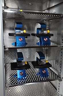
A key characteristic of time-lapse cytometers is their use of non heat-generating light sources such as light-emitting diodes. This allows a time-lapse cytometer to be placed inside a conventional cell culture incubator to facilitate continuous observation of cellular processes without heat building up inside the incubator.[6]
History

Hemocytometer
The early history of cytometry is closely associated with the development of the blood cell counting. Through the work of Karl von Vierordt, Louis-Charles Malassez, Karl Bürker and others blood cell concentration could by the late 19th century be accurately measured using a blood cell counting chamber, the hemocytometer, and an optical microscope.[7][8]
Until the 1950s the hemocytometer was the standard method to count blood cells.[9] In blood cell counting applications the hemocytometer has now been replaced by electronic cell counters. However, the hemocytometer is still being used to count cells in cell culture laboratories. Successively the manual task of counting, using a microscope, is taken over by small automated image cytometers.
Fluorescence microscope
As with most other microscopy development in the late 19th and early 20th century, the fluorescent microscope was pioneered by associates to Carl Zeiss AG in Jena, Germany. In 1904, Moritz von Rohr and August Köhler at Carl Zeiss constructed the first ultraviolet microscope. The intent of the microscope was to obtain higher optical resolution by using illumination with a shorter wavelength than visual light. However, they experienced difficulties with autofluorescence when observing biological material. Fortunately, Köhler saw the potential of fluorescence. This led him and the Carl Zeiss co-inventor of the ultramicroscope, Henry Siedentopf, to build a prototype of a specialized fluorescent microscope in 1908.[10][11] Unfortunately, the ultraviolet light source they used was somewhat dim and fluorescence microscopy remained impractical until Heinrich Lehmann in 1910 applied a much brighter light source and the ultraviolet filtering techniques developed by Robert Wood. Independently, Oskar Heimstädt worked along the same lines together with a competitor to Carl Zeiss; C Reichert, Optische Werke AG in Vienna, which today is a part of Leica Microsystems.[12][13][14]
Cytophotometry
By the early 1930s various firms manufactured ultraviolet fluorescent microscopes. The stage was set for cytometry to now go beyond the now established hemocytometer. At this time, Torbjörn Caspersson, working at the Karolinska Institute in Stockholm, developed a series of progressively more sophisticated instruments called cytophotometers. These instruments combined a fluorescent microscope with a spectrophotometer to quantify cellular nucleic acids and their relation to cell growth and function. Caspersson’s early apparatus now seems hopelessly primitive. But, even this primitive apparatus got results, and attracted the attention of other researchers. Many of the advances in analytical cytology from the 1940s and on-wards were made by people who made the pilgrimage to Stockholm.[3]
Pulse cytophotometry

The first attempts to automate cell counting were made around World War II. Gucker et al. builds a device to detect bacteria in aerosols.[15] Lagercrantz builds an automated cell counter based on microcopy[16] and identifies the difficulties in aligning cells to be individually counted using microscopy, as Moldavan had proposed in 1934.[17] Joseph and Wallace Coulter circumnavigates these difficulties by inventing the principle of using electrical impedance to count and size microscopic particles suspended in a fluid.[9][18] This principle is today known as the Coulter principle and was used in the automated blood cell counter released by Coulter Electronics in 1954. The “Coulter counter” was the first commercial flow cytometer.
During the 1960s Dittrich, Göhde and Kamentsky improves the design pioneered by Caspersson 30 years earlier. Dittrich and Göhde’s pulse cytophotometer was built around a Zeiss fluorescent microscope and commercialized as the ICP 11 by Partec GmbH in 1968. Kamentsky’s device was commercialized by Bio/Physics Systems Inc. as the Cytograph in 1970.[19][20] These devices were able to count cells, like the earlier Coulter counter. But more importantly, they were also capable of measuring cellular characteristics. However, these early cytophotometers where microscopy-based.[21]
Flow cytometry
In 1953 Crosland-Taylor published an unsuccessful attempt to count red blood cells using microscopy in which he solved the problem of aligning cells by using sheath fluid to hydrodynamicially focus the cells.[2] In the late 1960s, Van Dilla at Los Alamos National Laboratory built the first non microscopy-based cytophotometer. He did this by combining Crosland-Taylor's breakthrough with the fluorescent dyes originally developed for microscopy and a laser-based fluorescent detection system — the flow cytometer as we know it today.[22][23][24] Fulwyler, at Los Alamos as well, combines the Coulter principle with continuous inkjet printer technology to create the first cell sorter in 1965.[25] In 1973 Steinkamp and the team at Los Alamos follow up with a fluorescence-based cell sorter.[26]
In 1978, at the Conference of the American Engineering Foundation in Pensacola, Florida, the name pulse cytophotometry was changed to flow cytometry, a term which quickly became popular.[27] At that point pulse cytophotometry had evolved into the modern form of flow cytometry, pioneered by Van Dilla ten years earlier.
See also
References
- ↑ "International Society for Advancement of Cytometry". Retrieved 2013-03-31.
- 1 2 Crosland-Taylor, P. J. (1953). "A device for counting small particles suspended in a fluid through a tube". Nature. 171 (4340): 37–38. doi:10.1038/171037b0. PMID 13025472.
- 1 2 Shapiro H. (2004). "The Evolution of Cytometers". Cytometry Part A. 58A: 13–20. doi:10.1002/cyto.a.10111.
- ↑ Coutu, D. L.; Schroeder, T. (2013). "Probing cellular processes by long-term live imaging - historic problems and current solutions". Journal of Cell Science. 126 (Pt 17): 3805–15. doi:10.1242/jcs.118349. PMID 23943879.
- ↑ Pavillon, N; Kühn, J; Moratal, C; Jourdain, P; Depeursinge, C; Magistretti, P. J.; Marquet, P (2012). "Early cell death detection with digital holographic microscopy". PLoS ONE. 7 (1): e30912. doi:10.1371/journal.pone.0030912. PMC 3269420
 . PMID 22303471.
 - ↑ "Time-lapse Imaging Cytometers & Cell Incubator Microscopes". Phase Holographic Imaging AB. Retrieved 2013-09-10.
- ↑ Verso, M. L. (1964). "The Evolution of Blood-Counting Techniques". Med. Hist. 8 (2): 149–158. doi:10.1017/s0025727300029392. PMC 1033366. PMID 14139094.
- ↑ Verso, M. L. (1971). "Some nineteenth-century pioneers of haematology". Med. Hist. 15 (1): 55–67. doi:10.1017/s0025727300016124. PMC 1034115. PMID 4929622.
- 1 2 "The History of Flow Cytometry". Beckman-Coulter Inc. Retrieved 2013-03-31.
- ↑ Birgit Herden; Michael Zölffel (2008). "100 Years of Living Images" (PDF). Innovation. Carl Zeiss AG. 12 (21): 16–23.
- ↑ "History of Microscopes". Carl Zeiss AG. Retrieved 2013-03-31.
- ↑ Rusk, N. (2009). "The fluorescence microscope". Milstones in Light Microscopy. Nature Publishing Group.
- ↑ Heimstädt O. (1911). "Das Fluoreszenzmikroskop". Z. Wiss. Mikrosk. 28: 330–337.
- ↑ Rost, F. W. D. (1995). Fluorescent microscopy, volume II. Cambridge University Press. pp. 183–187.
- ↑ Gucker, F. T.; O’Konski, C. T.; Pickard, H. B.; Pitts, J. N. (1947). "A photoelectronic counter for colloidal particles". J Am Chem Soc. 69: 2422–2431. doi:10.1021/ja01202a053.
- ↑ Lagercrantz, C. (1948). "Photo-electric Counting of Individual Microscopic Plant and Animal Cells". Nature. 161 (4079): 25–26. doi:10.1038/161025b0.
- ↑ Moldavan, A. (1934). "Photo-Electric Technique for the Counting of Microscopical Cells". Science. 80 (2069): 188–189. doi:10.1126/science.80.2069.188. PMID 17817054.
- ↑ U.S. patent 2656508, Coulter W. H., "Means for Counting Particles Suspended in a Fluid.", issued 1953-10-20
- ↑ "Flow Museum". Partec GmbH. Retrieved 2013-08-24.
- ↑ DE 1815352, Wolfgang Dittrich & Wolfgang Göhde, "Flow-through Chamber for Photometers to Measure and Count Particles in a Dispersion Medium"
- ↑ Kamentsky, L. A.; Melamed, M. R.; Derman, H (1965). "Spectrophotometer: New instrument for ultrarapid cell analysis". Science. 150 (3696): 630–1. doi:10.1126/science.150.3696.630. PMID 5837105.
- ↑ Van Dilla, M. A.; Trujillo, T. T.; Mullaney, P. F.; Coulter, J. R. (1969). "Cell microfluorometry: A method for rapid fluorescence measurement". Science. 163 (3872): 1213–1214. doi:10.1126/science.163.3872.1213. PMID 5812751.
- ↑ "Cytometry Volume 10 – Los Alamos". Purdue University Cytometry Laboratories. Retrieved 2013-08-24.
- ↑ Robinson, J. P. (2009). "Cytometry – a Definitive History of the Early Days". In Sack, U.; Tárnok, A.; Rothe, G. Cellular Diagnostics. Basics, Methods and Clinical Applications of Flow. Karger. pp. 1–28.
- ↑ Fulwyler, M. J. (1965). "Electronic separation of biological cells by volume". Science. 150 (698): 910–911. doi:10.1126/science.150.3698.910. PMID 5891056.
- ↑ Steinkamp, J. A.; Fulwyler, M. J.; Coulter, J. R.; Hiebert, R. D.; Horney, J. L.; Mullancy, P. F. (1973). "A new multiparameter separator for microscopic particles and biological cells". The Review of scientific instruments. 44 (9): 1301–1310. doi:10.1063/1.1686375. PMID 4279087.
- ↑ "Partec Flow Museum". Partec GmbH. Retrieved 2013-08-25.
External links
- International Society for Advancement of Cytometry
- International Clinical Cytometry Society
- Cytometry Volume 10 by Purdue University Cytometry Laboratories
- Time-lapse Image Cytometry by Phase Holographic Imaging AB