Zygomatic bone
| Zygomatic bone | |
|---|---|
 Left zygomatic bone in situ. | |
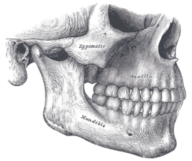 Side view of the teeth and jaws. (Zygomatic visible in center.) | |
| Details | |
| Identifiers | |
| Latin | os zygomaticum, zygoma |
| TA | A02.1.14.001 |
| FMA | 52747 |
In the human skull, the zygomatic bone (cheekbone or malar bone) is a paired bone which articulates with the maxilla, the temporal bone, the sphenoid bone and the frontal bone. It is situated at the upper and lateral part of the face and forms the prominence of the cheek, part of the lateral wall and floor of the orbit, and parts of the temporal and infratemporal fossa. It presents a malar and a temporal surface; four processes, the frontosphenoidal, orbital, maxillary, and temporal; and four borders.
Structure
Surfaces
The malar surface is convex and perforated near its center by a small aperture, the zygomaticofacial foramen, for the passage of the zygomaticofacial nerve and vessels; below this foramen is a slight elevation, which gives origin to the zygomaticus muscle.
The temporal surface, directed posteriorly and medially, is concave, presenting medially a rough, triangular area, for articulation with the maxilla (articular surface), and laterally a smooth, concave surface, the upper part of which forms the anterior boundary of the temporal fossa, the lower a part of the infratemporal fossa. Near the center of this surface is the zygomaticotemporal foramen for the transmission of the zygomaticotemporal nerve.
The orbital surface forms the lateral part and some of the inferior part of the bony orbit. The zygomatic nerve passes through the zygomatic-orbital foramen on this surface. The lateral palpebral ligament attaches to a small protuberance called the orbital tubercle.
Processes
Each zygomatic bone is diamond-shaped and composed of three processes with similarly named associated bony articulations: frontal, temporal, and maxillary. Each process of the zygomatic bone forms important structures of the skull.
The orbital surface of the frontal process of the zygomatic bone forms the anterior lateral orbital wall, with usually a small paired foramen, the zygomaticofacial foramen opening on its lateral surface. The temporal process of the zygomatic bone forms the zygomatic arch along with the zygomatic process of the temporal bone, with a paired zygomaticotemporal foramen present on the medial deep surface of the bone. The orbital surface of the maxillary process of the zygomatic bone forms a part of the infraorbital rim and a small part of the anterior part of the lateral orbital wall.[1]
Borders
The antero-superior or orbital border is smooth, concave, and forms a considerable part of the circumference of the orbit.
The antero-inferior or maxillary border is rough, and bevelled at the expense of its inner table, to articulate with the maxilla; near the orbital margin it gives origin to the Quadratus labii superioris.
The postero-superior or temporal border, curved like an italic letter f, is continuous above with the commencement of the temporal line, and below with the upper border of the zygomatic arch; the temporal fascia is attached to it.
The postero-inferior or zygomatic border affords attachment by its rough edge to the Masseter.
Articulations
The zygomatic articulates with four bones: the frontal, sphenoid, temporal, and maxilla.
Development
The zygomatic bone is generally described as ossifying from three centers— one for the malar and two for the orbital portion; these appear about the eighth week and fuse about the fifth month of fetal life.
Mall describes it as being ossified from one center which appears just beneath and to the lateral side of the orbit.
After birth, the bone is sometimes divided by a horizontal suture into an upper larger, and a lower smaller division.
In some quadrumana the zygomatic bone consisted of two parts, an orbital and a malar.
Society and culture
High cheekbones are pronounced zygomatic arches, causing the upper part of the cheeks to jut out and form a line cut into the sides of the face. High cheekbones, forming a symmetrical face shape, are very common in fashion models and are considered a beauty trait in both males and females.[2]
Etymology
The term zygomatic derives from the Greek Ζυγόμα zygoma meaning "yoke". The zygomatic bone is occasionally referred to as the zygoma, but this term may also refer to the zygomatic arch. The zygomatic is homologous to the jugal bone of other tetrapods.
Additional images
|


In other animals


In non-mammalian vertebrates, the zygomatic bone is referred to as the jugal bone, since these animals have no zygomatic arch. It is found in most reptiles, amphibians, and birds. It is connected to the quadratojugal and maxilla, as well as other bones, which may vary by species.
This bone is considered key in the determination of general traits of the skull, as in the case of creatures, such as dinosaurs in paleontology, whose entire skull has not been found. In coelacanths and early tetrapods the bone is relatively large. Here, it is a plate-like bone forming the lower margin of the orbit and much of the side of the face. In ray-finned fishes it is reduced or absent, and the entire cheek region is generally small. The bone is also absent in living amphibians.[3]
With the exception of turtles, the jugal bone in reptiles forms a relatively narrow bar separating the orbit from the inferior temporal fenestra, of which it may also form the lower boundary. The bone is similarly reduced in birds. In mammals, it takes on broadly the form seen in humans, with the bar between the orbit and fenestra vanishing entirely, and only the lower boundary of the fenestra remaining, as the zygomatic arch.[3]
See also
References
This article incorporates text in the public domain from the 20th edition of Gray's Anatomy (1918)
- ↑ Illustrated Anatomy of the Head and Neck, Fehrenbach and Herring, Elsevier, 2012, page 54.
- ↑ Sex and Society. Marshall Cavendish. September 2009. p. 91. ISBN 978-0-7614-7906-2. Retrieved 2 November 2012.
- 1 2 Romer, Alfred Sherwood; Parsons, Thomas S. (1977). The Vertebrate Body. Philadelphia, PA: Holt-Saunders International. pp. 217–241. ISBN 0-03-910284-X.
External links
| Wikimedia Commons has media related to Zygomatic bone. |
- Anatomy diagram: 34256.000-1 at Roche Lexicon - illustrated navigator, Elsevier
- Facial Bone Anatomy at eMedicine