Organelle
| Organelle | |
|---|---|
| Details | |
| Identifiers | |
| Latin | organella |
| Code | TH H1.00.01.0.00009 |
| TH | H1.00.01.0.00009 |
| FMA | 63832 |
| Cell biology | |
|---|---|
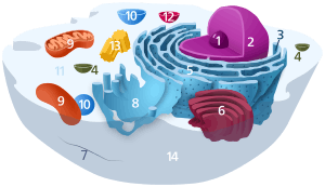
| The animal cell | |
|
Components of a typical animal cell:
|
In cell biology, an organelle /ɔːrɡəˈnɛl/ is a specialized subunit within a cell that has a specific function. Individual organelles are usually separately enclosed within their own lipid bilayers.
The name organelle comes from the idea that these structures are to cells what an organ is to the body (hence the name organelle, the suffix -elle being a diminutive). Organelles are identified by microscopy, and can also be purified by cell fractionation. There are many types of organelles, particularly in eukaryotic cells. While prokaryotes do not possess organelles per se, some do contain protein-based bacterial microcompartments, which are thought to act as primitive organelles.[1]
History and terminology
In biology organs are defined as confined functional units within an organism.[2] The analogy of bodily organs to microscopic cellular substructures is obvious, as from even early works, authors of respective textbooks rarely elaborate on the distinction between the two.
Credited as the first[3][4][5] to use a diminutive of organ (i.e., little organ) for cellular structures was German zoologist Karl August Möbius (1884), who used the term organula (plural of organulum, the diminutive of Latin organum).[6] In a footnote, which was published as a correction in the next issue of the journal, he justified his suggestion to call organs of unicellular organisms "organella" since they are only differently formed parts of one cell, in contrast to multicellular organs of multicellular organisms.
Types of organelles
While most cell biologists consider the term organelle to be synonymous with "cell compartment", other cell biologists choose to limit the term organelle to include only those that are DNA-containing, having originated from formerly autonomous microscopic organisms acquired via endosymbiosis.[7][8][9]
Under this definition, there would only be two broad classes of organelles (i.e. those that contain their own DNA, and have originated from endosymbiotic bacteria):
- mitochondria (in almost all eukaryotes)
- plastids[10] (e.g. in plants, algae, and some protists).
Other organelles are also suggested to have endosymbiotic origins, but do not contain their own DNA (notably the flagellum – see evolution of flagella).
Under the more restricted definition of membrane-bound structures, some parts of the cell do not qualify as organelles. Nevertheless, the use of organelle to refer to non-membrane bound structures such as ribosomes is common.[11] This has led some texts to delineate between membrane-bound and non-membrane bound organelles.[12] The non-membrane bound organelles, also called large biomolecular complexes, are large assemblies of macromolecules that carry out particular and specialized functions, but they lack membrane boundaries. Such cell structures include:
- large RNA and protein complexes: ribosome, spliceosome, vault
- large protein complexes: proteasome, DNA polymerase III holoenzyme, RNA polymerase II holoenzyme, symmetric viral capsids, complex of GroEL and GroES; membrane protein complexes: photosystem I, ATP synthase
- large DNA and protein complexes: nucleosome
- centriole and microtubule-organizing center (MTOC)
- cytoskeleton
- flagellum
- Nucleolus
- Stress granule
- Germ cell granule
- Neuronal transport granule
Eukaryotic organelles
Eukaryotic cells are structurally complex, and by definition are organized, in part, by interior compartments that are themselves enclosed by lipid membranes that resemble the outermost cell membrane. The larger organelles, such as the nucleus and vacuoles, are easily visible with the light microscope. They were among the first biological discoveries made after the invention of the microscope.
Not all eukaryotic cells have each of the organelles listed below. Exceptional organisms have cells that do not include some organelles that might otherwise be considered universal to eukaryotes (such as mitochondria).[13] There are also occasional exceptions to the number of membranes surrounding organelles, listed in the tables below (e.g., some that are listed as double-membrane are sometimes found with single or triple membranes). In addition, the number of individual organelles of each type found in a given cell varies depending upon the function of that cell.
| Organelle | Main function | Structure | Organisms | Notes |
|---|---|---|---|---|
| chloroplast (plastid) | photosynthesis, traps energy from sunlight | double-membrane compartment | plants, protists (rare kleptoplastic organisms) | has own DNA; theorized to be engulfed by the ancestral eukaryotic cell (endosymbiosis) |
| endoplasmic reticulum | translation and folding of new proteins (rough endoplasmic reticulum), expression of lipids (smooth endoplasmic reticulum) | single-membrane compartment | all eukaryotes | rough endoplasmic reticulum is covered with ribosomes, has folds that are flat sacs; smooth endoplasmic reticulum has folds that are tubular |
| Flagellum | locomotion, sensory | some eukaryotes | ||
| Golgi apparatus | sorting, packaging, processing and modification of proteins | single-membrane compartment | all eukaryotes | cis-face (convex) nearest to rough endoplasmic reticulum; trans-face (concave) farthest from rough endoplasmic reticulum |
| mitochondria | energy production from the oxidation of glucose substances and the release of adenosine triphosphate | double-membrane compartment | most eukaryotes | has own DNA; theorized to be engulfed by an ancestral eukaryotic cell (endosymbiosis) |
| vacuole | storage, transportation, helps maintain homeostasis | single-membrane compartment | eukaryotes | |
| nucleus | DNA maintenance, controls all activities of the cell, RNA transcription | double-membrane compartment | all eukaryotes | contains bulk of genome |
Mitochondria and chloroplasts, which have double-membranes and their own DNA, are believed to have originated from incompletely consumed or invading prokaryotic organisms, which were adopted as a part of the invaded cell. This idea is supported in the Endosymbiotic theory.
| Organelle/Macromolecule | Main function | Structure | Organisms |
|---|---|---|---|
| acrosome | helps spermatozoa fuse with ovum | single-membrane compartment | many animals |
| autophagosome | vesicle that sequesters cytoplasmic material and organelles for degradation | double-membrane compartment | all eukaryotes |
| centriole | anchor for cytoskeleton, organizes cell division by forming spindle fibers | Microtubule protein | animals |
| cilium | movement in or of external medium; "critical developmental signaling pathway".[14] | Microtubule protein | animals, protists, few plants |
| eyespot apparatus | detects light, allowing phototaxis to take place | green algae and other unicellular photosynthetic organisms such as euglenids | |
| glycosome | carries out glycolysis | single-membrane compartment | Some protozoa, such as Trypanosomes. |
| glyoxysome | conversion of fat into sugars | single-membrane compartment | plants |
| hydrogenosome | energy & hydrogen production | double-membrane compartment | a few unicellular eukaryotes |
| lysosome | breakdown of large molecules (e.g., proteins + polysaccharides) | single-membrane compartment | animals |
| melanosome | pigment storage | single-membrane compartment | animals |
| mitosome | probably plays a role in Iron-sulfur cluster (Fe-S) assembly | double-membrane compartment | a few unicellular eukaryotes that lack mitochondria |
| myofibril | myocyte contraction | bundled filaments | animals |
| nematocyst | stinging | coiled hollow tubule | Cnidarians |
| nucleolus | pre-ribosome production | protein-DNA-RNA | most eukaryotes |
| parenthesome | not characterized | not characterized | fungi |
| peroxisome | breakdown of metabolic hydrogen peroxide | single-membrane compartment | all eukaryotes |
| proteasome | degradation of unneeded or damaged proteins by proteolysis | very large protein complex | All eukaryotes, all archaea, some bacteria |
| ribosome (80S) | translation of RNA into proteins | RNA-protein | all eukaryotes |
| vesicle | material transport | single-membrane compartment | all eukaryotes |
| Stress granule | mRNA storage[15] | membraneless
(mRNP complexes) |
Most eukaryotes |
Other related structures:
Prokaryotic organelles
Prokaryotes are not as structurally complex as eukaryotes, and were once thought not to have any internal structures enclosed by lipid membranes. In the past, they were often viewed as having little internal organization, but slowly, details are emerging about prokaryotic internal structures. An early false turn was the idea developed in the 1970s that bacteria might contain membrane folds termed mesosomes, but these were later shown to be artifacts produced by the chemicals used to prepare the cells for electron microscopy.[17]
However, more recent research has revealed that at least some prokaryotes have microcompartments such as carboxysomes. These subcellular compartments are 100–200 nm in diameter and are enclosed by a shell of proteins.[1] Even more striking is the description of membrane-bound magnetosomes in bacteria, reported in 2006,[18][19] as well as the nucleus-like structures of the Planctomycetes that are surrounded by lipid membranes, reported in 2005.[20]
| Organelle/Macromolecule | Main function | Structure | Organisms |
|---|---|---|---|
| carboxysome | carbon fixation | protein-shell compartment | some bacteria |
| chlorosome | photosynthesis | light harvesting complex | green sulfur bacteria |
| flagellum | movement in external medium | protein filament | some prokaryotes and eukaryotes |
| magnetosome | magnetic orientation | inorganic crystal, lipid membrane | magnetotactic bacteria |
| nucleoid | DNA maintenance, transcription to RNA | DNA-protein | prokaryotes |
| plasmid | DNA exchange | circular DNA | some bacteria |
| ribosome (70S) | translation of RNA into proteins | RNA-protein | bacteria and archaea |
| thylakoid | photosynthesis | photosystem proteins and pigments | mostly cyanobacteria |
| mesosomes | functions of Golgi bodies, centrioles, etc. | small irregular shaped organelle containing ribosomes | present in most prokaryotic cells |
| Pilus | Adhesion to other cells for conjugation or to a solid substrate to create motile forces. | a hair-like appendage sticking out (though partially embedded into) the plasma membrane | prokaryotic cells |
Proteins and organelles
The function of a protein is closely correlated with the organelle in which it resides. Some methods were proposed for predicting the organelle in which an uncharacterized protein is located according to its amino acid composition[21][22] and some methods were based on pseudo amino acid composition.[23][24][25][26]
See also
- CoRR Hypothesis
- Ejectosome
- Endosymbiotic theory
- Organelle biogenesis
- Membrane vesicle trafficking
- Host-pathogen interface
References
- 1 2 Kerfeld, C. A.; Sawaya, M. R; Tanaka, S; Nguyen, C. V.; Phillips, M; Beeby, M; Yeates, T. O. (5 August 2005). "Protein structures forming the shell of primitive bacterial organelles.". Science. 309 (5736): 936–8. Bibcode:2005Sci...309..936K. doi:10.1126/science.1113397. PMID 16081736.
- ↑ Lynsey Peterson (2010-04-17). "Mastering the Parts of a Cell". Lesson Planet. Retrieved 2010-04-19.
- ↑ Bütschli, O. (1888). Dr. H. G. Bronn's Klassen u. Ordnungen des Thier-Reichs wissenschaftlich dargestellt in Wort und Bild. Erster Band. Protozoa. Dritte Abtheilung: Infusoria und System der Radiolaria. p. 1412.
Die Vacuolen sind demnach in strengem Sinne keine beständigen Organe oder O r g a n u l a (wie Möbius die Organe der Einzelligen im Gegensatz zu denen der Vielzelligen zu nennen vorschlug).
- ↑ Amer. Naturalist. 23, 1889, p. 183: "It may possibly be of advantage to use the word organula here instead of organ, following a suggestion by Möbius. Functionally differentiated multicellular aggregates in multicellular forms or metazoa are in this sense organs, while, for functionally differentiated portions of unicellular organisms or for such differentiated portions of the unicellular germ-elements of metazoa, the diminutive organula is appropriate." Cited after: Oxford English Dictionary online, entry for "organelle".
- ↑ Robin, Ch. (1891). Journal de l'anatomie et de la physiologie normales et pathologiques de l'homme et des animaux. F. Alcan.
- ↑ Möbius, K. (September 1884). "Das Sterben der einzelligen und der vielzelligen Tiere. Vergleichend betrachtet". Biologisches Centralblatt. 4 (13, 14): 389–392, 448.
Während die Fortpflanzungszellen der vielzelligen Tiere unthätig fortleben bis sie sich loslösen, wandern und entwickeln, treten die einzelligen Tiere auch durch die an der Fortpflanzung beteiligten Leibesmasse in Verkehr mit der Außenwelt und viele bilden sich dafür auch besondere Organula". Footnote on p. 448: "Die Organe der Heteroplastiden bestehen aus vereinigten Zellen. Da die Organe der Monoplastiden nur verschieden ausgebildete Teile e i n e r Zelle sind schlage ich vor, sie „Organula“ zu nennen
- ↑ Keeling, Pj; Archibald, Jm (2008). "Organelle evolution: what's in a name?". Current Biology. 18 (8): R345–7. doi:10.1016/j.cub.2008.02.065. PMID 18430636.
- ↑ Imanian B, Carpenter KJ, Keeling PJ (2007). "Mitochondrial genome of a tertiary endosymbiont retains genes for electron transport proteins". The Journal of eukaryotic microbiology. 54 (2): 146–53. doi:10.1111/j.1550-7408.2007.00245.x. PMID 17403155.
- ↑ Mullins, Christopher (2004). "Theory of Organelle Biogenesis: A Historical Perspective". The Biogenesis of Cellular Organelles. Springer Science+Business Media, National Institutes of Health. ISBN 0-306-47990-7.
- ↑ Hogan, C. Michael (2010). Deoxyribonucleic acid. Encyclopedia of Earth. National Council for Science and the Environment. S. Draggan and C. Cleveland (eds.). Washington DC
- ↑ Campbell and Reece, Biology 6th edition, Benjamin Cummings, 2002
- ↑ Cormack, David H. (1984) Introduction to Histology, Lippincott, ISBN 0397521146
- ↑ Fahey RC, Newton GL, Arrack B, Overdank-Bogart T, Baley S (1984). "Entamoeba histolytica: a eukaryote without glutathione metabolism". Science. 224 (4644): 70–72. Bibcode:1984Sci...224...70F. doi:10.1126/science.6322306. PMID 6322306.
- ↑ Badano, Jose L.; Norimasa Mitsuma; Phil L. Beales; Nicholas Katsanis (September 2006). "The Ciliopathies: An Emerging Class of Human Genetic Disorders". Annual Review of Genomics and Human Genetics. 7: 125–148. doi:10.1146/annurev.genom.7.080505.115610. PMID 16722803.
- ↑ Anderson, Paul; Kedersha, Nancy (2008-03-01). "Stress granules: the Tao of RNA triage". Trends in Biochemical Sciences. 33 (3): 141–150. doi:10.1016/j.tibs.2007.12.003. PMID 18291657.
- ↑ Tsai Y, Sawaya MR, Cannon GC, Cai F, Williams EB, Heinhorst S, Kerfeld CA, Yeates TO (2007). "Structural Analysis of CsoS1A and the Protein Shell of the Halothiobacillus neapolitanus Carboxysome". PLoS Biology. 5 (6): e144. doi:10.1371/journal.pbio.0050144. PMC 1872035
 . PMID 17518518.
 - ↑ Ryter A (1988). "Contribution of new cryomethods to a better knowledge of bacterial anatomy". Ann. Inst. Pasteur Microbiol. 139 (1): 33–44. doi:10.1016/0769-2609(88)90095-6. PMID 3289587.
- ↑ Komeili A, Li Z, Newman DK, Jensen GJ (2006). "Magnetosomes are cell membrane invaginations organized by the actin-like protein MamK". Science. 311 (5758): 242–5. Bibcode:2006Sci...311..242K. doi:10.1126/science.1123231. PMID 16373532.
- ↑ Scheffel A, Gruska M, Faivre D, Linaroudis A, Plitzko JM, Schüler D (2006). "An acidic protein aligns magnetosomes along a filamentous structure in magnetotactic bacteria". Nature. 440 (7080): 110–4. Bibcode:2006Natur.440..110S. doi:10.1038/nature04382. PMID 16299495.
- ↑ Fuerst JA (2005). "Intracellular compartmentation in planctomycetes". Annu. Rev. Microbiol. 59: 299–328. doi:10.1146/annurev.micro.59.030804.121258. PMID 15910279.
- ↑ Cedano, J.; Aloy, P.; P'erez-Pons, J. A.; Querol, E. (1997). "Relation between amino acid composition and cellular location of proteins". J. Mol. Biol. 266 (3): 594–600. doi:10.1006/jmbi.1996.0804. PMID 9067612.
- ↑ Chou, K. C.; Elrod, D. W. (1999). "Protein subcellular location prediction". Protein Engineering. 12 (2): 107–118. doi:10.1093/protein/12.2.107. PMID 10195282.
- ↑ Chou, KC (2001). "Prediction of protein cellular attributes using pseudo-amino acid composition". Proteins. 43 (3): 246–55. doi:10.1002/prot.1035. PMID 11288174.
- ↑ Mundra, P.; Kumar, M.; Kumar, K. K.; Jayaraman, V. K.; Kulkarni, B. D. (2007). "Using pseudo amino acid composition to predict protein subnuclear localization: Approached with PSSM". Pattern Recognition Letters. 28 (13): 1610–1615. doi:10.1016/j.patrec.2007.04.001.
- ↑ Du, P.; Cao, S.; Li, Y. (2009). "SubChlo: predicting protein subchloroplast locations with pseudo-amino acid composition and the evidence-theoretic K-nearest neighbor (ET-KNN) algorithm". Journal of Theoretical Biology. 261 (2): 330–335. doi:10.1016/j.jtbi.2009.08.004.
- ↑ Li, F. M.; Li, Q. Z. (2008). "Predicting protein subcellular location using Chou's pseudo amino acid composition and improved hybrid approach". Protein & Peptide Letters. 15 (6): 612–616. doi:10.2174/092986608784966930. PMID 18680458.
External links
| Library resources about Organelle |