Anterior compartment of leg
| Anterior compartment of leg | |
|---|---|
 | |
| Details | |
| Artery | anterior tibial artery |
| Nerve | deep fibular nerve |
| Identifiers | |
| Latin | Compartimentum cruris anterius |
| TA | A04.7.01.005 |
| FMA | 45163 |
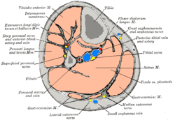
The anterior compartment of the leg is a fascial compartment of the lower limb. It contains muscles that produce dorsiflexion and participate in inversion and eversion of the foot, as well as vascular and nervous elements including the anterior tibial artery and veins, and the deep fibular nerve.
Muscles
The muscles of the compartment are:[1]
- tibialis anterior
- extensor hallucis longus
- extensor digitorum longus
- fibularis tertius (peroneus tertius)
| Muscle | Proximal Attachment | Distal Attachment | Innervation | Main Action |
|---|---|---|---|---|
| Tibialis anterior | Lateral condyle and superior half of lateral surface of tibia and interosseous membrane | Medial and inferior surfaces of medial cuneiform and base of 1st metatarsal | Deep fibular nerve (L4, L5) | Dorsiflexes ankle and inverts foot |
| Extensor digitorum longus | Lateral condyle of tibia and superior three quarters of medial surface of fibula and interosseous membrane | Middle and distal phalanges of lateral four digits | Extends lateral four digits and dorsiflexes ankle | |
| Extensor hallucis longus | Middle part of anterior surface of fibula and interosseous membrane | Dorsal aspect of base of distal phalanx of great toe (hallux) | Extends great toe and dorsiflexes ankle | |
| Fibularis tertius | Inferior third of anterior surface of fibula and interosseous membrane | Dorsum of base of 5th metatarsal | Dorsiflexes ankle and aids in eversion of foot |
Function
The compartment contains muscles that are dorsiflexors and participate in inversion and eversion of the foot.[2]
Innervation and blood supply
The anterior compartment of the leg is supplied by the deep fibular nerve (deep peroneal nerve), a branch of the common fibular nerve. The nerve contains axons from the L4, L5, and S1 spinal nerves.
Blood for the compartment is supplied by the anterior tibial artery, which runs between the tibialis anterior and extensor digitorum longus muscles. When the artery crosses the flexor retinaculum, it changes its name to dorsal artery of foot.[3]
See also
Notes and references
- ↑ Moore, Dally, and Agur (2014). Moore Clinically Oriented Anatomy, Table 5.10, p 591.
- ↑ antlegdorsalfoot at The Anatomy Lesson by Wesley Norman (Georgetown University)
- ↑ "Vessels and lympatic drainage of the lower limb". Retrieved 11 January 2015.