Acetabular fracture
| Acetabular fracture | |
|---|---|
|
Acetabular fracture as seen on plain X-ray | |
| Classification and external resources | |
| ICD-10 | S32.4 |
| ICD-9-CM | xxx |

Fractures of the acetabulum occur when the head of the femur is driven into the pelvis. This is caused either by a blow on the side or by a blow in the front of the knee, usually in a dashboard injury when the femur also may be fractured.[1]
Acetabulum in Latin means a cup that holds vinegar. (Collins English Dictionary). It is a cavity situated on the outer surface of Innominate bone. Innominate bone is confluence of 3 bones, the ilium, ischium and pubis. Acetabulum and ball of femur form the hip joint.
Fractures of the acetabulum in young individuals usually occur due to high energy injury like vehicular accident or fall from the height. While in old individuals, who have osteoporotic (fragile) bones, can occur due to trivial fall.
The energy is transferred from the femoral head to the acetabulum resulting in the fracture. The type of the fracture depends on the position of the limb and the direction of the force causing variety of fractures
The credit of classifying these fractures and giving guidelines for surgical management goes to French surgeons, Robertt Judet, Jean Judet and Emile Letournel, who in their landmark paper in 1964, described the column concept of the innominate bone, mechanism of injury, classification and surgical management of these complex injuries. According to them, the acetabulum is situated between two columns of innominate bone, namely anterior Ilio-pubic column and posterior Ilio-ischial column. They classified these fractures into elementary (simple two part) and associated (complex three or more part) fractures.
Anatomy


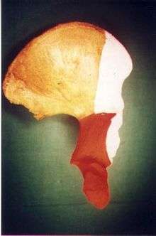
To understand fracture pattern it is essential to have minimum three x-ray views;
- · Pelvis with both hips antero posterior view
This view shows six important landmarks of the acetabulum, viz;
- 1. Pelvic brim
- 2. Ilio ischial line
- 3. Tear drop
- 4. Anterior wall
- 5. Posterior wall
- 6. Weight bearing dome
2· Iliac oblique view
Shows whole of ilium, the posterior column and the anterior wall
3.· Obturator oblique view
Shows the anterior column and the posterior wall
Use of CT scan with 3-D reconstruction of images has made understanding of these fractures easier.
Judet-Letournel classification
Elementary fractures
Posterior wall
Posterior column
Anterior wall
Anterior column
Transverse
Associated fractures
T shape
Posterior column + Posterior wall
Transverse + posterior wall
Anterior column + posterior hemi transverse
Combined both column fractures
Elementary fractures
Posterior wall fracture


This is the most common variety of fracture
Cause: Occurs due to dash board injury. When a person travelling in a vehicle involved in head-on collision, the force applied over the flexed knee travels along the femur bone to the head of femur breaking the posterior wall of the acetabulum. The head of the femur is dislocated outside the joint.
Associated injury: The broken bone pieces or the dislocated head of the femur may injure the important Sciatic nerve causing paralysis of the foot; this may or may not recover depending on the extent of injury to the nerve.
The posterior wall fragment may be one large piece, or multiple pieces, and may be associated with impaction of the bone
How to diagnose: Best seen in obturator oblique view
CT scan helps in identifying impaction of bone pieces and if there are pieces in the joint
MRI may be done to identify the extent of injury to the sciatic nerve
Treatment: if the femur head is dislocated, it should be reduced as soon as possible, to prevent damage to its blood supply. This is preferably done under anaesthesia, following which, leg is kept pulled by applying traction to prevent joint from dislocating.
The final management depends on the size of the fragment(s), stability and congruence of the joint.
In some cases traction for six to eight weeks may be the only treatment required
If the fragments do not fall into place, or if there are bone pieces in the joint, or if the joint is unstable, surgical fixation using screw(s) and plate(s) is performed
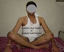
Post-surgery treatment: depending on the stability achieved, the person may be allowed standing and walking with help of support for about six to eight weeks.
Full function may return in about three months.
Complications: Sciatic nerve injury and stoppage of blood supply to femoral head at the time of accident or during surgery may occur. Deep vein thrombosis and pulmonary embolism are other complications that may occur in any type of injury to the acetabulum.
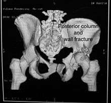
Posterior column fracture
Cause: as with posterior wall injury, this also occurs due to dash board injury.
Associated injury: There could be associated fracture of the posterior wall making this associated variety of fracture. The broken bone piece may injure the important Sciatic nerve causing paralysis of the foot, may or may not recover depending on the extent of injury to the nerve.
How to diagnose: Best seen in iliac oblique and obturator oblique views
CT scan helps in identifying impaction of bone pieces and if there are pieces in the joint
Treatment: if the femur head is dislocated, it should be reduced as soon as possible, to prevent damage to its blood supply. This is preferably done under anaesthesia, following which, leg is kept pulled by applying traction to prevent joint from dislocating.
The final management depends on the size of the fragment(s), stability and congruence of the joint.
In some cases traction for six to eight weeks may be the only treatment required
If the fragments do not fall into place, or if there are bone pieces in the joint, or if the joint is unstable, surgical fixation using screw(s) and plate(s) is performed
Post-surgery treatment: depending on the stability achieved, the person may be allowed standing and walking with help of support for about six to eight weeks.
Full function may return in about three months.
Complications: Sciatic nerve injury and stoppage of blood supply to femoral head at the time of accident or during surgery may occur. Deep vein thrombosis and pulmonary embolism are other complications that may occur in any type of injury to the acetabulum.
Anterior wall fracture

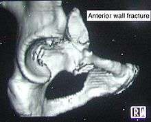

Not a very common injury
Cause: occurs when the force applied from the side. On the greater trochanter of the femur bone, as in fall on the side or being hit on the side
Associated injury: usually none
How to diagnose: Best seen in iliac oblique view
CT scan helps in identifying impaction of bone pieces and if there are pieces in the joint
Treatment: if the femur head is dislocated, it should be reduced as soon as possible, to prevent damage to its blood supply. This is preferably done under anaesthesia, following which, leg is kept pulled by applying traction to prevent joint from dislocating.
The final management depends on the size of the fragment(s), stability and congruence of the joint.
In some cases traction for six to eight weeks may be the only treatment required
If the fragments do not fall into place, or if there are bone pieces in the joint, or if the joint is unstable, surgical fixation using screw(s) and plate(s) is performed
Post-surgery treatment: depending on the stability achieved, the person may be allowed standing and walking with help of support for about six to eight weeks.
Full function may return in about three months.
Complications: Stoppage of blood supply to femoral head at the time of accident or during surgery may occur. Deep vein thrombosis and pulmonary embolism are other complications that may occur in any type of injury to the acetabulum.
Anterior column fracture

Cause: Occurs when the force applied from the side. On the greater trochanter of the femur bone, as in fall on the side or being hit on the side
Types: Depending on the location, the fractures are described as very low, low, intermediate and high anterior column fracture.
Associated injury: There could be associated fracture of the posterior column making this associated variety of fracture named as anterior with posterior hemi transverse fracture.
How to diagnose: Best seen in Obturator oblique view

CT scan helps in identifying impaction of bone pieces and if there are pieces in the joint
Treatment: if the femur head is dislocated, it should be reduced as soon as possible, to prevent damage to its blood supply. This is preferably done under anaesthesia, following which, leg is kept pulled by applying traction to prevent joint from dislocating.
The final management depends on the size of the fragment(s), stability and congruence of the joint.
In some cases traction for six to eight weeks may be the only treatment required
If the fragments do not fall into place, or if there are bone pieces in the joint, or if the joint is unstable, surgical fixation using screw(s) and plate(s) is performed
Post-surgery treatment: depending on the stability achieved, the person may be allowed standing and walking with help of support for about six to eight weeks.
Full function may return in about three months.
Complications: Stoppage of blood supply to femoral head at the time of accident or during surgery may occur. Deep vein thrombosis and pulmonary embolism are other complications that may occur in any type of injury to the acetabulum.
Transverse fracture
In this variety of fracture, the innominate bone is broken such that upper part consists of Ilium with weight bearing dome and lower part consists of Ischium and Pubic bones. This is a two part fracture. Though both columns are broken, still the weight bearing dome is still attached to main ilium and hence it is not a true both column fracture.
Types: Depending on the level at which the fracture line passes in relation to weight bearing area, the transverse fracture is further subdivided into;
1. Infra tectal: below the weight bearing dome
2. Juxta tectal: just at the level of the weight bearing dome
3. Trans tectal: passing through the weight bearing dome
(Tectum = any roof like structure in the body: Collins English dictionary)
Cause: Occurs when the force applied from the side. On the greater trochanter of the femur bone, as in fall on the side or being hit on the side. This force may be combined with dash board injury as well.
Associated injury: There could be associated fracture of the posterior wall making this associated variety of fracture.
How to diagnose: Best seen in Antero posterior view and Iliac and obturator oblique views
In CT scan the characteristic feature is that the fracture line runs from front to back
CT scan helps in identifying impaction of bone pieces and if there are pieces in the joint
Treatment: if the femur head is dislocated, it should be reduced as soon as possible, to prevent damage to its blood supply. This is preferably done under anaesthesia, following which, leg is kept pulled by applying traction to prevent joint from dislocating.
The final management depends on the size of the fragment(s), stability and congruence of the joint.
In some cases traction for six to eight weeks may be the only treatment required
If the fragments do not fall into place, or if there are bone pieces in the joint, or if the joint is unstable, surgical fixation using screw(s) and plate(s) is performed
Post-surgery treatment: depending on the stability achieved, the person may be allowed standing and walking with help of support for about six to eight weeks.
Full function may return in about three months.
Complications: Injury to the femoral head, stoppage of blood supply to femoral head at the time of accident or during surgery may occur. Sciatic nerve may get damaged at the time of injury or during surgery. Deep vein thrombosis and pulmonary embolism are other complications that may occur in any type of injury to the acetabulum.
Associated fractures
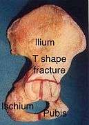
T shape fracture
When a transverse fracture also had a vertical fracture line, it is called a T shape fracture. Here the innominate bone is broken in such a way that all three parts of it, the Ilium, the Ischium and the Pubis are separated from one another. This is a three part fracture. Though both columns are broken, still the weight bearing dome is still attached to main ilium and hence it is not a true both column fracture.
This fracture may be associated with fracture through the posterior wall as well, making it more complex
Cause: Occurs when the force applied from the side. On the greater trochanter of the femur bone, as in fall on the side or being hit on the side. This force may be combined with dash board injury as well.

Associated injury: Injury to the sciatic nerve
How to diagnose: Best seen in Antero posterior view and Iliac and obturator oblique views
CT scan helps in identifying impaction of bone pieces and if there are pieces in the joint
Treatment: if the femur head is dislocated, it should be reduced as soon as possible, to prevent damage to its blood supply. This is preferably done under anaesthesia, following which, leg is kept pulled by applying traction to prevent joint from dislocating.
The final management depends on the size of the fragment(s), stability and congruence of the joint.

In some cases traction for six to eight weeks may be the only treatment required
If the fragments do not fall into place, or if there are bone pieces in the joint, or if the joint is unstable, surgical fixation using screw(s) and plate(s) is performed
Post-surgery treatment: depending on the stability achieved, the person may be allowed standing and walking with help of support for about six to eight weeks.
Full function may return in about three months.
Complications: Injury to the femoral head, stoppage of blood supply to femoral head at the time of accident or during surgery may occur. Sciatic nerve may get damaged at the time of injury or during surgery. Deep vein thrombosis and pulmonary embolism are other complications that may occur in any type of injury to the acetabulum.
Posterior column with posterior wall / Transverse with posterior wall Fractures

These fractures are extension of elementary fractures. With involvement of posterior wall, the difficulty in treatment increases. These fractures are rarely amenable to non-surgical treatment. Due to posterior wall fracture, the hip is usually dislocated posteriorly, requiring immediate reduction of dislocation and surgical reconstruction after few days.
Cause: Posterior column with posterior wall fracture occurs due to dash board injury, while transverse fracture with posterior wall fracture occurs due to combined dash board injury and direct injury to the hip from the side.
Associated injury: to the sciatic nerve may occur
How to diagnose: Antero posterior view may give clue to these injuries, Judet views and CT scan help in knowing the extent of injury
Treatment: if the femur head is dislocated, it should be reduced as soon as possible, to prevent damage to its blood supply. This is preferably done under anaesthesia, following which, leg is kept pulled by applying traction to prevent joint from dislocating.
The final management depends on the size of the fragment(s), stability and congruence of the joint.
In some cases traction for six to eight weeks may be the only treatment required
If the fragments do not fall into place, or if there are bone pieces in the joint, or if the joint is unstable, surgical fixation using screw(s) and plate(s) is performed
Post-surgery treatment: depending on the stability achieved, the person may be allowed standing and walking with help of support for about six to eight weeks.
Full function may return in about three months.
Complications: Injury to the femoral head, stoppage of blood supply to femoral head at the time of accident or during surgery may occur. Sciatic nerve may get damaged at the time of injury or during surgery. Deep vein thrombosis and pulmonary embolism are other complications that may occur in any type of injury to the acetabulum.
Anterior with posterior hemi transverse fractures
In this variety of fracture, the posterior or ilio ischial column is broken as a transverse fracture while the anterior or ilio pubic column is broken into multiple pieces. Part of the weight bearing dome in this variety of fracture is still attached to that part of iliac wing which is forms part of sacro iliac joint. This type of injury has to be differentiated from both column fracture, where in the weight bearing dome is a floating piece not attached directly to bone forming sacro iliac joint
Cause: Combination of forces acting on the hip though the femoral head
Associated injury: Sciatic nerve, may be femoral nerve or vessels
How to diagnose: all three x-ray views plus CT scan is a must for diagnosis and management of this complex injury
Treatment: like any other acetabular fracture, if the femoral head is dislocated out of the socket, early reduction into socket is a priority. Non operative treatment rarely gives satisfactory result. If the patient is unfit to undergo major surgery due to any reason, longitudinal traction to achieve secondary congruence of hip may help to restore hip function, though partially.
Surgical management is ideal. The choice of approach rests with the surgeon, but going from front, or anterior approach is must. The posterior injury may be tacked with anterior approach by experienced surgeon.
Post-surgery treatment: depending on the stability achieved, the person may be allowed standing and walking with help of support for about six to eight weeks.
Full function may return in about three months.
Complications: Injury to the femoral head, stoppage of blood supply to femoral head at the time of accident or during surgery may occur. Sciatic nerve may get damaged at the time of injury or during surgery. Anterior approach may cause damage to lateral cutaneous nerve of the thigh, injury to femoral nerve of femoral vessels may occur.
Deep vein thrombosis and pulmonary embolism are other complications that may occur in any type of injury to the acetabulum.
Combined both column fractures
These are the most complex injuries. Here the weight bearing roof or dome of the acetabulum is a floating piece. This adds to complexity of management.


Cause: Combination of forces acting on the hip though the femoral head
Associated injury: Sciatic nerve, may be femoral nerve or vessels
How to diagnose: all three x-ray views plus CT scan is a must for diagnosis and management of this complex injury
Treatment: like any other acetabular fracture, if the femoral head is dislocated out of the socket, early reduction into socket is a priority. Non operative treatment rarely gives satisfactory result. If the patient is unfit to undergo major surgery due to any reason, longitudinal traction to achieve secondary congruence of hip may help to restore hip function, though partially.
Surgical management is ideal. The choice of approach rests with the surgeon, but going from front, or anterior approach is preferred. The posterior injury may be tacked with anterior approach by experienced surgeon.
Post-surgery treatment: depending on the stability achieved, the person may be allowed standing and walking with help of support for about six to eight weeks.
Full function may return in about three months.
Complications: Injury to the femoral head, stoppage of blood supply to femoral head at the time of accident or during surgery may occur. Sciatic nerve may get damaged at the time of injury or during surgery. Anterior approach may cause damage to lateral cutaneous nerve of the thigh, injury to femoral nerve of femoral vessels may occur.
Deep vein thrombosis and pulmonary embolism are other complications that may occur in any type of injury to the acetabulum.
Principles of management
At the site of injury: After stabilizing an injured person and resuscitation, quick examination is done to check injury to vital organs.
If one suspects injury to the hip, it is imperative to immobilse the limb using some kind of support to prevent movements of the injured limb to prevent further damage
A trained paramedic may be able to diagnose hip dislocation by noticing the position of the injured limb. It is essential to document status of nerves and vessels before starting any treatment to protect oneself from litigation
On arrival at the hospital, trained trauma surgeon will assess the patient and prescribe necessary tests including x-rays as described earlier.
Non-surgical management consists of reducing the dislocated joint by maneuver under anaesthesia and applying traction to the limb to maintain position of joint and fractured bones. If non surgical management is preferred it may require six weeks to 3 months for recovery.
Surgical management
The surgical management requires high degree of training and well equipped centre. It should be carried out by experienced surgical team to get best results
The principles laid down for management are;
- · Anatomic reduction of the fractured fragments
- · Stable fixation
- · Congruent joint
- · Early mobilization
- · Delayed weight bearing
Innominate bone is a flat bone with many curves. In most part the bone is thick enough and has broad surfaces that are amenable to primary fixation using lag screw(s) and to neutralize forces across the bone one needs to add plate(s) on the surface of the fractured fragments for it to heal without deformity.
Before surgery, patient needs tests to check fitness for surgery
Anaesthesia : the surgery may be performed either under regional anaesthesia or general anaesthesia
Surgical approaches. Following are the common approaches;
- · Kocher Langenbeck approach for posterior injuries
- · Ili inguinal, Ilio femoral of modified stoppa’s approach for anterior or combined injuries
Implants : normally lag screws and reconstruction plates are preferred implants
Post operative management: would involve initial period or bed rest, followed by mobilsation by trained therapist
Total time to recover may be up to 3 months
Patterns of fracture
Tile's classification of acetabular fracture:
- I - Simple fracture, anterior or posterior wall column
- II - Transverse fracture
- III - T - Type fracture involving two columns
- IV - Both columns fractures, floating acetabulum
-

Axial CT image (viewed on bone windows) of a complex comminuted left acetabular fracture involving both anterior and posterior columns.
-

Fracture of the acetabulum
References
| 1. | Matta JM, Anderson LM, Epstein HC, Hendricks P. Fractures of acetabulum: a retrospective analysis. Clin Orthop 1986; 205:230. |
| 2. | Rowe CR, Lowell JD. Prognosis of fractures of acetabulum. J Bone Joint Surg [Am] 1961; 43A: 30 - - 59. |
| 3. | Tile M. Fractures of pelvis and acetabulum. Baltimore; Williams & Wilkins. 1984 |
| 4. | Letournel E. Acetabular fractures, classification and management. Clin Orthop 1980; 151: 81-106. |
| 5. | Pennal GF, Davidson J, Garside H, Lewis J. Results of treatment of acetabular fractures. Clin Orthop 1980; 151: 115 - 123. |
| 6. | Tile M, Schatzker J. Rationale of operative fracture care. Berlin, Heidelberg, New York; Springer -Verlag. 1987. |
- ↑ Solomon, APLEY'S TRAUMA AND ORTHOPAEDICS, EIGHT EDITION,