ATP5E


| View/Edit Human | |||
| Mitochondrial ATP synthase epsilon chain | |||||||||
|---|---|---|---|---|---|---|---|---|---|
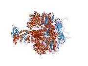
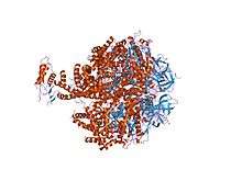 ground state structure of f1-atpase from bovine heart mitochondria (bovine f1-atpase crystallised in the absence of azide) | |||||||||
| Identifiers | |||||||||
| Symbol | ATP-synt_Eps | ||||||||
| Pfam | PF04627 | ||||||||
| InterPro | IPR006721 | ||||||||
| SCOP | 1e79 | ||||||||
| SUPERFAMILY | 1e79 | ||||||||
| |||||||||
ATP synthase subunit epsilon, mitochondrial is an enzyme that in humans is encoded by the ATP5E gene.[2][3]
This gene encodes a subunit of mitochondrial ATP synthase. Mitochondrial ATP synthase catalyzes ATP synthesis, utilizing an electrochemical gradient of protons across the inner membrane during oxidative phosphorylation. ATP synthase is composed of two linked multi-subunit complexes: the soluble catalytic core, F1, and the membrane-spanning component, Fo, comprising the proton channel. The catalytic portion of mitochondrial ATP synthase consists of 5 different subunits (alpha, beta, gamma, delta, and epsilon) assembled with a stoichiometry of 3 alpha, 3 beta, and a single representative of the other 3. The proton channel consists of three main subunits (a, b, c). This gene encodes the epsilon subunit of the catalytic core. Two pseudogenes of this gene are located on chromosomes 4 and 13.[3]
Structure
The ATP5E gene, located on the q arm of chromosome 20 in position 13.32, is made up of 3 exons and is 3,690 base pairs in length.[3] The ATP5A1 protein weighs 5.7 kDa and is composed of 51 amino acids.[4][5] The protein is a subunit of the F1Fo ATPase, also known as Complex V, which consists of 14 nuclear and 2 mitochondrial -encoded subunits. As an alpha subunit, ATP5A1 is contained within the catalytic F1 portion of the complex and acts as a regulatory subunit that inhibits the binding of ADP, preventing wasteful ATP production.[3] The nomenclature of the enzyme has a long history. The F1 fraction derives its name from the term "Fraction 1" and Fo (written as a subscript letter "o", not "zero") derives its name from being the binding fraction for oligomycin, a type of naturally-derived antibiotic that is able to inhibit the Fo unit of ATP synthase.[6][7] The F1 particle is large and can be seen in the transmission electron microscope by negative staining.[8] These are particles of 9 nm diameter that pepper the inner mitochondrial membrane. They were originally called elementary particles and were thought to contain the entire respiratory apparatus of the mitochondrion, but, through a long series of experiments, Efraim Racker and his colleagues (who first isolated the F1 particle in 1961) were able to show that this particle is correlated with ATPase activity in uncoupled mitochondria and with the ATPase activity in submitochondrial particles created by exposing mitochondria to ultrasound. This ATPase activity was further associated with the creation of ATP by a long series of experiments in many laboratories.
Function
Mitochondrial membrane ATP synthase (F1Fo ATP synthase or Complex V) produces ATP from ADP in the presence of a proton gradient across the membrane which is generated by electron transport complexes of the respiratory chain. F-type ATPases consist of two structural domains, F1 - containing the extramembraneous catalytic core, and Fo - containing the membrane proton channel, linked together by a central stalk and a peripheral stalk. During catalysis, ATP synthesis in the catalytic domain of F1 is coupled via a rotary mechanism of the central stalk subunits to proton translocation. Part of the complex F1 domain and of the central stalk which is part of the complex rotary element. Rotation of the central stalk against the surrounding alpha3beta3 subunits leads to hydrolysis of ATP in three separate catalytic sites on the beta subunits (By similarity).[9]
The epsilon subunit is located in the stalk region of the F1 complex, and acts as an inhibitor of the ATPase catalytic core. The epsilon subunit can assume two conformations, contracted and extended, where the latter inhibits ATP hydrolysis. The conformation of the epsilon subunit is determined by the direction of rotation of the gamma subunit, and possibly by the presence of ADP. The epsilon subunit is thought to become extended in the presence of ADP, thereby acting as a safety lock to prevent wasteful ATP hydrolysis.[10]
Clinical significance
Mutations in the ATP5E gene cause mitochondrial complex V deficiency, nuclear 3 (MC5DN3), a mitochondrial disorder with heterogeneous clinical manifestations including dysmorphic features, psychomotor retardation, hypotonia, growth retardation, cardiomyopathy, enlarged liver, hypoplastic kidneys and elevated lactate levels in urine, plasma and cerebrospinal fluid.[11]
Reduced expression of ATP5E is significantly associated with the diagnosis of Papillary Thyroid Cancer and may serve as an early tumor marker of the disease.[12] Papillary Thyroid Cancer is the most common type of thyroid cancer,[13] representing 75 percent to 85 percent of all thyroid cancer cases.[14] It occurs more frequently in women and presents in the 20–55 year age group. It is also the predominant cancer type in children with thyroid cancer, and in patients with thyroid cancer who have had previous radiation to the head and neck.[15]
References
- ↑ "Human PubMed Reference:".
- ↑ Tu Q, Yu L, Zhang P, Zhang M, Zhang H, Jiang J, Chen C, Zhao S (Jun 2000). "Cloning, characterization and mapping of the human ATP5E gene, identification of pseudogene ATP5EP1, and definition of the ATP5E motif". Biochem J. 347 (1): 17–21. doi:10.1042/0264-6021:3470017. PMC 1220925
 . PMID 10727396.
 - 1 2 3 4 "Entrez Gene: ATP5E ATP synthase, H+ transporting, mitochondrial F1 complex, epsilon subunit".
- ↑ Zong NC, Li H, Li H, Lam MP, Jimenez RC, Kim CS, Deng N, Kim AK, Choi JH, Zelaya I, Liem D, Meyer D, Odeberg J, Fang C, Lu HJ, Xu T, Weiss J, Duan H, Uhlen M, Yates JR, Apweiler R, Ge J, Hermjakob H, Ping P (Oct 2013). "Integration of cardiac proteome biology and medicine by a specialized knowledgebase". Circulation Research. 113 (9): 1043–53. doi:10.1161/CIRCRESAHA.113.301151. PMC 4076475. PMID 23965338.
- ↑ "ATP synthase subunit epsilon, mitochondrial". Cardiac Organellar Protein Atlas Knowledgebase (COPaKB).
- ↑ Kagawa Y, Racker E (1966). "Partial resolution of the enzymes catalyzing oxidative phosphorylation. 8. Properties of a factor conferring oligomycin sensitivity on mitochondrial adenosine triphosphatase.". Journal of Biological Chemistry. 241: 2461–2466. PMID 4223640.
- ↑ Mccarty RE (November 1992). "A plant biochemist's view of H+
-ATPases and ATP synthases". J. Exp. Biol. 172 (Pt 1): 431–441. PMID 9874753. - ↑ Fernández-Morán H, Oda T, Blair PV, Green DE (July 1964). "A macromolecular repeating unit of mitochondrial structure and function. Correlated electron microscopic and biochemical studies of isolated mitochondria and submitochondrial particles of beef heart muscle". J. Cell Biol. 22 (1): 63–100. doi:10.1083/jcb.22.1.63. PMC 2106494. PMID 14195622.
- ↑ "ATP synthase subunit epsilon, mitochondrial". UniProt. The UniProt Consortium.
- ↑ Feniouk BA, Junge W (September 2005). "Regulation of the F0F1-ATP synthase: the conformation of subunit epsilon might be determined by directionality of subunit gamma rotation". FEBS Lett. 579 (23): 5114–8. doi:10.1016/j.febslet.2005.08.030. PMID 16154570.
- ↑ "ATP5E". Genetics Home Resource. NCBI.
- ↑ Hurtado-López, LM; Fernández-Ramírez, F; Martínez-Peñafiel, E; Carrillo Ruiz, JD; Herrera González, NE (16 June 2015). "Molecular Analysis by Gene Expression of Mitochondrial ATPase Subunits in Papillary Thyroid Cancer: Is ATP5E Transcript a Possible Early Tumor Marker?". Medical science monitor : international medical journal of experimental and clinical research. 21: 1745–51. doi:10.12659/MSM.893597. PMID 26079849.
- ↑ Hu MI, Vassilopoulou-Sellin R, Lustig R, Lamont JP "Thyroid and Parathyroid Cancers" in Pazdur R, Wagman LD, Camphausen KA, Hoskins WJ (Eds) Cancer Management: A Multidisciplinary Approach. 11 ed. 2008.
- ↑ Chapter 20 in: Mitchell, Richard Sheppard; Kumar, Vinay; Abbas, Abul K; Fausto, Nelson. Robbins Basic Pathology. Philadelphia: Saunders. ISBN 1-4160-2973-7. 8th edition.
- ↑ Dinets A, Hulchiy M, Sofiadis A, Ghaderi M, Höög A, Larsson C, Zedenius J (2012). "Clinical, Genetic and Immunohistochemical Characterization of 70 Ukrainian Adult Cases with Post-Chornobyl Papillary Thyroid Carcinoma". Eur J Endocrinol. 166: 1049–60. doi:10.1530/EJE-12-0144. PMC 3361791. PMID 22457234.
Further reading
- Viñas O, Powell SJ, Runswick MJ, et al. (1990). "The epsilon-subunit of ATP synthase from bovine heart mitochondria. Complementary DNA sequence, expression in bovine tissues and evidence of homologous sequences in man and rat.". Biochem. J. 265 (2): 321–6. PMC 1136890. PMID 2137333.
- Elston T, Wang H, Oster G (1998). "Energy transduction in ATP synthase.". Nature. 391 (6666): 510–3. doi:10.1038/35185. PMID 9461222.
- Wang H, Oster G (1998). "Energy transduction in the F1 motor of ATP synthase.". Nature. 396 (6708): 279–82. doi:10.1038/24409. PMID 9834036.
- Hu RM, Han ZG, Song HD, et al. (2000). "Gene expression profiling in the human hypothalamus-pituitary-adrenal axis and full-length cDNA cloning.". Proc. Natl. Acad. Sci. U.S.A. 97 (17): 9543–8. doi:10.1073/pnas.160270997. PMC 16901. PMID 10931946.
- Gross C, Kussmann S, Hehr A, et al. (2001). "Epsilon subunit gene of F(1)F(0)-ATP synthase (ATP5E) on human chromosome 20q13.2→q13.3 localizes between D20S171 and GNAS1.". Cytogenet. Cell Genet. 91 (1-4): 105–6. doi:10.1159/000056828. PMID 11173840.
- Deloukas P, Matthews LH, Ashurst J, et al. (2002). "The DNA sequence and comparative analysis of human chromosome 20.". Nature. 414 (6866): 865–71. doi:10.1038/414865a. PMID 11780052.
- Strausberg RL, Feingold EA, Grouse LH, et al. (2003). "Generation and initial analysis of more than 15,000 full-length human and mouse cDNA sequences.". Proc. Natl. Acad. Sci. U.S.A. 99 (26): 16899–903. doi:10.1073/pnas.242603899. PMC 139241. PMID 12477932.
- Cross RL (2004). "Molecular motors: turning the ATP motor.". Nature. 427 (6973): 407–8. doi:10.1038/427407b. PMID 14749816.
- Gerhard DS, Wagner L, Feingold EA, et al. (2004). "The status, quality, and expansion of the NIH full-length cDNA project: the Mammalian Gene Collection (MGC).". Genome Res. 14 (10B): 2121–7. doi:10.1101/gr.2596504. PMC 528928. PMID 15489334.
PDB gallery | ||||||||||
|---|---|---|---|---|---|---|---|---|---|---|
| ||||||||||


This article incorporates text from the United States National Library of Medicine, which is in the public domain.
This article incorporates text from the public domain Pfam and InterPro IPR006721